Journal of Forensic Medicine ›› 2022, Vol. 38 ›› Issue (4): 452-458.DOI: 10.12116/j.issn.1004-5619.2020.401215
• Original Article • Previous Articles Next Articles
Hai-yan LI1( ), Wen-gang LIU1, Shi-hai CUI1, Guang-long HE2, Peng XIA3, Li-juan HE1, Wen-le LÜ1
Received:2020-12-30
Online:2022-08-25
Published:2022-08-28
CLC Number:
Hai-yan LI, Wen-gang LIU, Shi-hai CUI, Guang-long HE, Peng XIA, Li-juan HE, Wen-le LÜ. Reconstruction and Quantitative Evaluation of Blunt Injury Cases by Finite Element Method[J]. Journal of Forensic Medicine, 2022, 38(4): 452-458.
Add to citation manager EndNote|Ris|BibTeX
URL: http://www.fyxzz.cn/EN/10.12116/j.issn.1004-5619.2020.401215
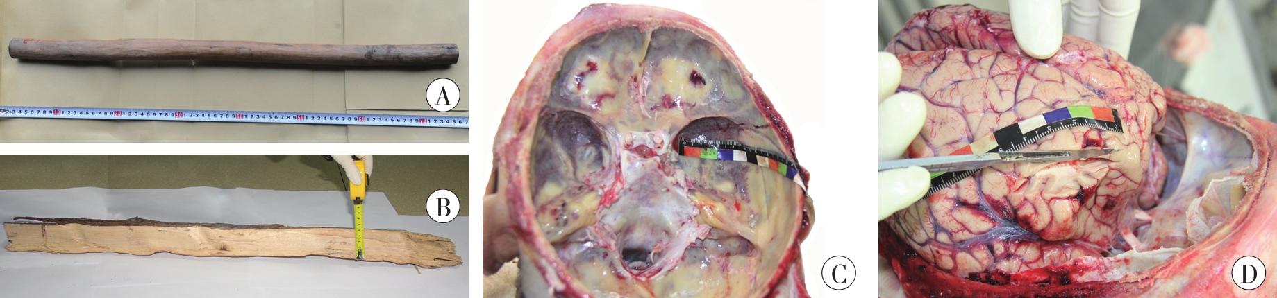
Fig. 1 Injury tools and craniocerebral injuries of case 1
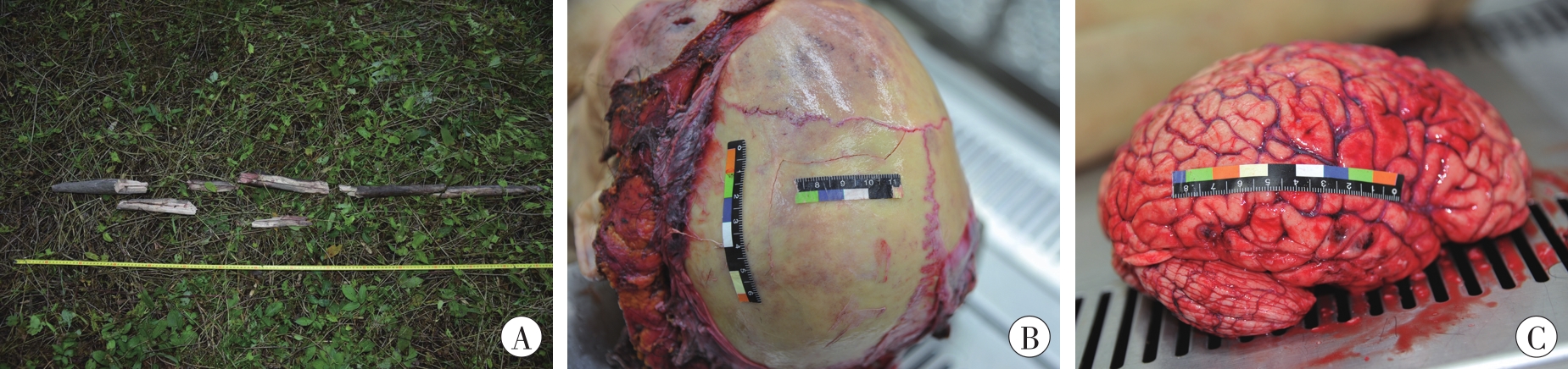
Fig. 2 Injury tools and craniocerebral injuries of case 2
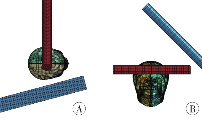
Fig. 3 Simulation settings of case 1 and case 2
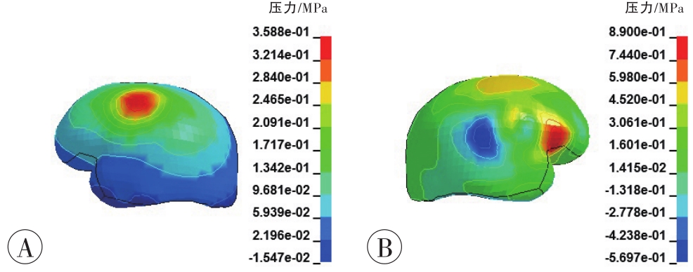
Fig. 4 The contours of the intracranial pressure of brain gray matter in case 1
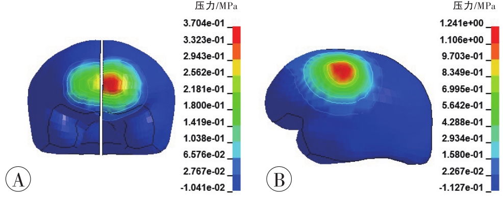
Fig. 5 The contours of the intracranial pressure of brain gray matter in case 2
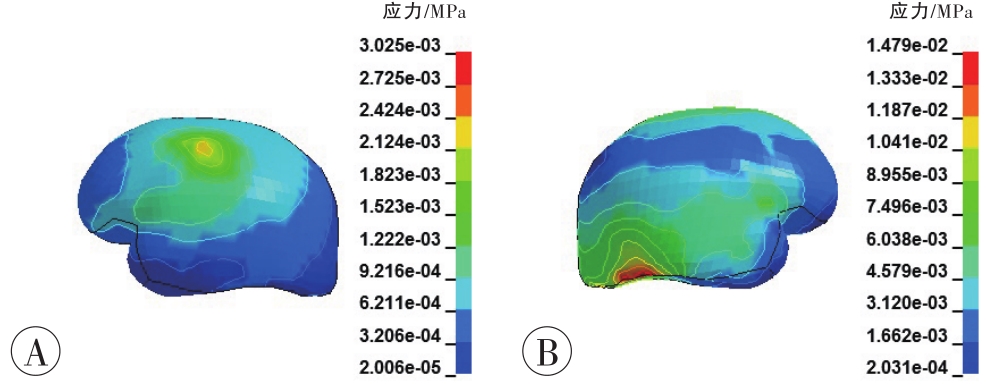
Fig. 6 The contours of the von Mises stress of brain tissue in case 1
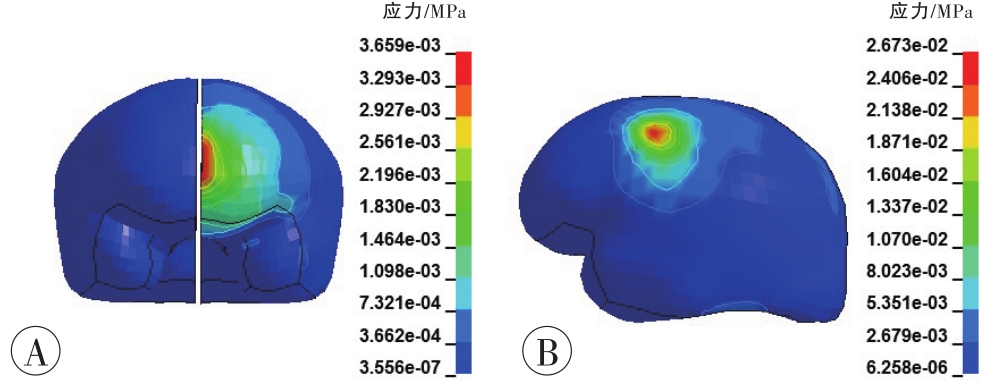
Fig. 7 The contours of the von Mises stress of brain tissue in case 2
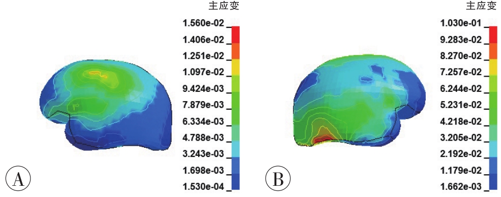
Fig. 8 The contours of the maximum principal strain of brain tissue in case 1
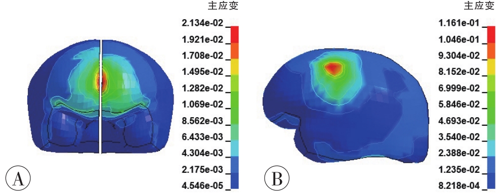
Fig. 9 The contours of the maximum principal strain of brain tissue in case 2
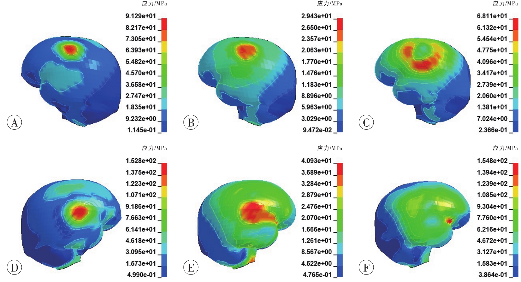
Fig. 10 The contours of the von Mises stress of skull in case 1
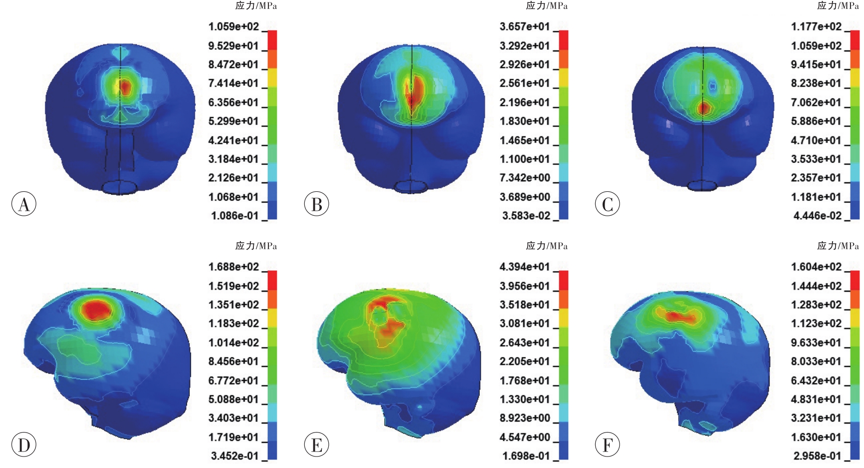
Fig. 11 The contours of the von Mises stress of skull in case 2
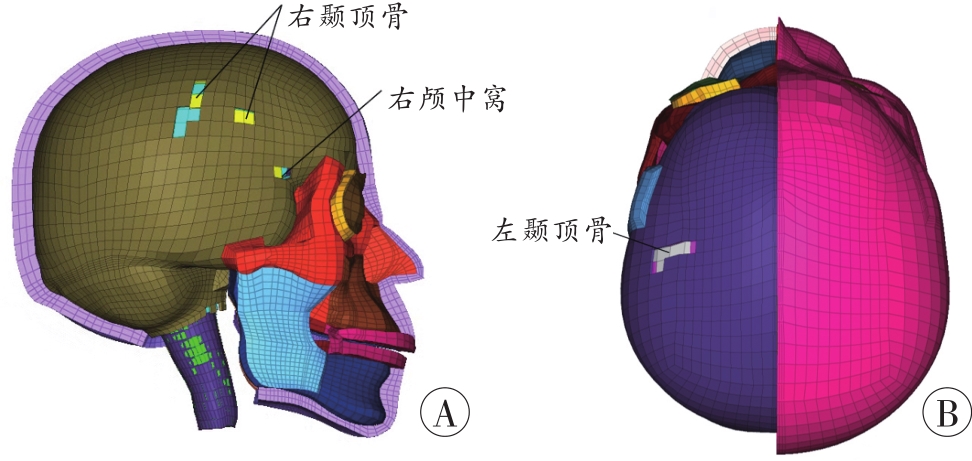
Fig. 12 Plastic strain of skull
| 1 | 李宗壮,吴俊蓉. 颅脑损伤的法医学鉴定研究[J].科技创新导报,2015,12(9):15. doi:10.16660/j.cnki.1674-098x.2015.09.091 . |
| LI Z Z, WU J R. Research on forensic identification of craniocerebral injury[J]. Keji Chuangxin Daobao,2015,12(9):15. | |
| 2 | MOTHERWAY J, DOORLY M C, CURTIS M, et al. Head impact biomechanics simulations: A forensic tool for reconstructing head injury?[J]. Leg Med (Tokyo),2009,11(S1):S220-S222. doi:10.1016/ |
| j.legalmed.2009.01.072. | |
| 3 | LI K, WANG J, LIU S, et al. Biomechanical behavior of brain injury caused by sticks using finite element model and Hybrid-Ⅲ testing[J]. Chin J Traumatol,2015,18(2):65-73. doi:10.1016/j.cjtee.2015.03.004 . |
| 4 | ZHAO W, RUAN S, LI H, et al. Development and validation of a 5th percentile human head finite element model based on the Chinese population[J]. Int J Veh Saf,2012,6(2):91-109. doi:10.1504/ijvs. 2012.049024 . |
| 5 | 赵玮. 基于碰撞生物力学的颅脑损伤机理及损伤评估方法研究[D].天津:天津科技大学,2012. |
| ZHAO W. Biomechanical exploration of brain injury mechanisms and injury assessment method evaluations[D]. Tianjin: Tianjin University of Science & Technology,2012. | |
| 6 | 夏哲浩. 基于ANSYS/LS-DYNA切削土壤与木材的数值模拟与实验分析[D].北京:北京林业大学,2016. |
| XIA Z H. Based on ANSYS-LS-DYNA the numerical simulation and experimental research of cutting soil and wood[D]. Beijing: Beijing Forestry University,2016. | |
| 7 | 潘建宇. 钝器载荷下颅骨骨折的有限元法评价研究[D].天津:天津科技大学,2020. |
| PAN J Y. Evaluation of skull fracture under blunt loading by finite element method[D]. Tianjin: Tianjin University of Science & Technology,2020. | |
| 8 | 包朝胜,黄必朝. 头部木质棍棒伤的法医鉴定[J].法医学杂志,1998,14(3):159. |
| BAO C S, HUANG B C. Forensic identification of head injury by club[J]. Fayixue Zazhi,1998,14(3):159. | |
| 9 | WARD C C, THOMPSON R B. The development of a detailed finite element brain model[C]// Proceeding of 19th Stapp Car Crash Conference. Society of Automotive Engineers,1975:641-674. |
| 10 | MILLER R T, MARGULIES S S, LEONI M, et al. Finite element modeling approaches for predicting injury in an experimental model of severe diffuse axonal injury[C]// Proceeding of 42nd Stapp Car Crash Conference, Society of Automotive Engineers,1998:2798-2810. |
| 11 | TAKHOUNTS E G, CRAIG M J, MOORHOUSE K, et al. Development of brain injury criteria (BrIC)[J]. Stapp Car Crash J,2013,57:243-266. doi:10.4271/2013-22-0010 . |
| 12 | 毛征宇,李泽民,牛文鑫,等. 不同载荷作用下头部生物力学响应仿真分析[J].医用生物力学,2016,31(6):532-539,547. doi:10.3871/j.1004-7220.2016.06.532 . |
| MAO Z Y, LI Z M, NIU W X, et al. The simulation analysis on biomechanical responses of human head under different loading conditions[J]. Yi-yong Shengwu Lixue,2016,31(6):532-539,547. | |
| 13 | BAUMGARTNER D, WILLINGER R, SHEW-CHENKO N, et al. Tolerance limits for mild traumatic brain injury derived from numerical head impact replication[C]// Proceedings of the 2001 International IRCOBI Conference on the Biomechanics of Impact, Isle of Man, UK:2001. |
| 14 | GALBRAITH J A, THIBAULT L E, MATTESON D R. Mechanical and electrical responses of the squid giant axon to simple elongation[J]. J Biomech Eng,1993,115(1):13-22. doi:10.1115/1.2895464 . |
| 15 | 毛少平. CT、MRI在弥漫性轴索损伤法医学鉴定中应用价值分析[J].中外医疗,2018,37(27):193-195. |
| doi:10.16662/j.cnki.1674-0742.2018.27.193.MAO S P. Application value analysis of CT and MRI in forensic identification of diffuse axonal injury[J]. Zhongwai Yiliao,2018,37(27):193-195. | |
| 16 | 林国威,左人宇,陆钊,等. 斜抛撑在基坑支护中控制土体应力与结构受力特点研究[J].土工基础,2015,29(4):13-18. |
| LIN G W, ZUO R Y, LU Z, et al. Structural capacity of the inclined supporting system and soil stress in the deep excavation[J]. Tugong Jichu,2015,29(4):13-18. | |
| 17 | 李晓东,左太阳. 颅骨内板凹陷性骨折的CT诊断(附4例分析)[J].实用放射学杂志,2005,21(1):97,110. |
| LI X D, ZUO T Y. CT diagnosis of depressed fracture in the skull inner lamina (An analysis of 4 cases)[J]. Shiyong Fangshexue Zazhi,2005,21(1):97,110. |
| [1] | Yong ZENG, Dong-hua ZOU, Ying FAN, Qing XU, Lu-yang TAO, Yi-jiu CHEN, Zheng-dong LI. Research Progress and Forensic Application of Human Vascular Finite Element Modeling and Biomechanics [J]. Journal of Forensic Medicine, 2023, 39(5): 471-477. |
| [2] | Hong-xia HAO, Jie-min CHEN, Rong-rong WANG, Xiao-ying YU, Meng WANG, Zhi-lu ZHOU, Yan-liang SHENG, Wen-tao XIA. The Value of VR-PVEP in Objective Evaluation of Monocular Refractive Visual Impairment [J]. Journal of Forensic Medicine, 2023, 39(4): 382-387. |
| [3] | Jie BAI, Jing SUN, Xiao-guang CHENG, Fan LIU, Hua LIU, Xu WANG. Construction and Application of Rib Fracture Diagnosis Model Based on YOLOv3 Algorithm [J]. Journal of Forensic Medicine, 2023, 39(4): 343-349. |
| [4] | Fei FAN, Juan WU, Zhen-hua DENG. Application Progress of Objective Audiological Detection Techniques in Forensic Clinical Medicine [J]. Journal of Forensic Medicine, 2023, 39(4): 360-366. |
| [5] | Jian XIANG, Xu WANG, Li-li YU, Kang-jia JIN, Ying-kai YANG. Objective Assessment of Visual Field Defects Caused by Optic Chiasm and Its Posterior Visual Pathway Injury [J]. Journal of Forensic Medicine, 2023, 39(4): 350-359. |
| [6] | Zhang-ming GAO, Jing-yu SHI, Hao ZENG, Xue-jun ZHANG. Rapid Determination of Bucinnazine in Blood by UPLC-MS/MS [J]. Journal of Forensic Medicine, 2023, 39(4): 388-392. |
| [7] | Yu-qi CAO, Yan SHI, Ping XIANG, Yin-long GUO. Research Progress on Machine Learning Assisted Non-Targeted Screening Strategy for Identification of Fentanyl Analogs [J]. Journal of Forensic Medicine, 2023, 39(4): 406-416. |
| [8] | Xu WANG. Legal Theories, Disability Models and Principles of Disability Assessment [J]. Journal of Forensic Medicine, 2023, 39(4): 329-336. |
| [9] | Hao-yang WANG, Jian WU, Qian ZHANG, Xin-feng MIN, Xiu-yan LIU, Yin-long GUO. Structural Analysis and Characterization of 4-F-α-PVP Analog 4-F-3-Methyl-α- PVP Hydrochloride [J]. Journal of Forensic Medicine, 2023, 39(2): 144-150. |
| [10] | Hang CHEN, Jing HU, Zheng QIAO, Hong-xiao DENG, Min LÜ, Wei LIU. Research Progress on Biological Matrix Reference Materials in Forensic Toxicology [J]. Journal of Forensic Medicine, 2023, 39(2): 176-185. |
| [11] | Jiao-jiao JI, Duo-qi XU, Ping XIANG, Hui YAN, Min SHEN. Analysis of Forty-Two Psychoactive Substances in a Single Hair by Micro-Segmental Technique [J]. Journal of Forensic Medicine, 2023, 39(2): 151-160. |
| [12] | Zhong-ping CHENG, Yan-fei LIU, Xing-min XU, Yao-nan MO. Progress in the Application of Magnetic Nanoparticles in Forensic Trace Analysis [J]. Journal of Forensic Medicine, 2023, 39(2): 168-175. |
| [13] | Jiang-wei YAN, Jun-hong SUN, Hong-xing WANG, Zhi-wen WEI, Xiang-jie GUO, Ji LI, Cai-rong GAO, Geng-qian ZHANG, Xin-hua LIANG, Qiang-qiang ZHANG, Hong-wei WANG, Si-jin LI, Ying-yuan WANG, Ke-ming YUN. Exploration and Practice of the “One Combination, Two Highlights, Three Combinations, Four in One” Innovative Talents Training Mode in Forensic Medicine [J]. Journal of Forensic Medicine, 2023, 39(2): 193-199. |
| [14] | Dong GAO, Pei-pei ZHUO, Dong TIAN, Dan RAN, Qing XIA, Wen-tao XIA. Correlation between Elbow Flexor Muscle Strength and Needle Electromyography Parameters after Musculocutaneous Nerve Injury [J]. Journal of Forensic Medicine, 2023, 39(2): 137-143. |
| [15] | Hao-tian MA, Hua-ye XIONG, Ye LU, Bing LI, Jiang-hua LAI. Analysis of the Development Trend in Forensic Odontology Based on CiteSpace [J]. Journal of Forensic Medicine, 2023, 39(1): 18-26. |
| Viewed | ||||||
|
Full text |
|
|||||
|
Abstract |
|
|||||