导航切换
Journal of Forensic Medicine
Home
About
About Journal
Editorial Board
Abstracting & Indexing
Awards
Authors
Guide to Authors
Writing Requirement
Ethics Standard
Subscribe
Advertisement
Contact Us
中文
Topics
Virtual Forensic Anthropology
DENG Zhen-hua
Default
Latest
Most Read
Please wait a minute...
For Selected:
Download Citations
EndNote
Ris
BibTeX
Toggle Thumbnails
Select
Journal of Forensic Medicine 2020, 36 (
5
): 595-604. DOI:
10.12116/j.issn.1004-5619.2020.05.001
Abstract
(
286
)
PDF(pc)
(806KB)(
1069
)
Knowledge map
Save
Related Articles
|
Metrics
|
Comments
(
0
)
Select
Research Progress of Adult Age Determination with Imaging Methods
FAN Fei, ZHAN Meng-jun, DAI Xin-hua , et al.
Journal of Forensic Medicine 2020, 36 (
5
): 605-613. DOI:
10.12116/j.issn.1004-5619.2020.05.002
Abstract
(
343
)
PDF(pc)
(687KB)(
1109
)
Knowledge map
Save
Adult age determination plays an important role in individual identification, criminal investigation and social welfare. The most popular adult age determination indicators are pubic symphysis, iliac auricular surface, costal cartilage, cranial sutures, teeth, laryngeal cartilage, etc. In recent years, with the progress of CT imaging and 3D reconstruction technology, the adult age determination study gradually has transferred from a time-consuming general observation of bones with complex pre-processing in the past to the non-destructive, convenient, time-saving and easy to store image analysis technology. To explore more accurate, rapid and convenient adult age determination methods, multiple imaging methods and artificial intelligence have been applied in adult age determination. This paper reviews the common methods and research progress of adult age determination at home and abroad, infers the development direction of adult age determination, in order to provide reference for the improvement and optimization of forensic adult age determination.
Related Articles
|
Metrics
|
Comments
(
0
)
Select
Research Progress and Prospect of Facial Reconstruction in Forensic Science
ZHAO Jia-min , CHU Guang, MOU Qing-nan , et al.
Journal of Forensic Medicine 2020, 36 (
5
): 614-621. DOI:
10.12116/j.issn.1004-5619.2020.05.003
Abstract
(
584
)
PDF(pc)
(686KB)(
1450
)
Knowledge map
Save
Facial reconstruction is a way to recover facial morphology by restoring soft tissues based on unidentified skulls using the knowledge of anatomy, anthropology, aesthetics, and computer science. It is applied in forensic science, oral plastic surgery and archeology, and especially plays an important role in the identification of the origin of the unknown corpses in forensic science. Facial reconstruction is the supplementary means of identification when other approaches (such as DNA comparison, imaging matching, dental records comparison, etc.) cannot identify individual identity. Facial soft tissue thickness (FSTT) is the basis of facial reconstruction and with the development of imaging and computer science, the techniques for measuring FSTT are improving rapidly and many related researches have appeared. This paper summarizes the application of facial reconstruction in forensic science, the accuracy of different methods and the research progress of this field to provide reference to this field.
Related Articles
|
Metrics
|
Comments
(
0
)
Select
Comparison of Three CNN Models Applied in Bone Age Assessment of Pelvic Radiographs of Adolescents
PENG Li- qin , WAN Lei , WANG Mao- wen, et al.
Journal of Forensic Medicine 2020, 36 (
5
): 622-630. DOI:
10.12116/j.issn.1004-5619.2020.05.004
Abstract
(
320
)
PDF(pc)
(1683KB)(
1147
)
Knowledge map
Save
Objective To compare the performance of three deep-learning models (VGG19, Inception-V3 and Inception-ResNet-V2) in automatic bone age assessment based on pelvic X-ray radiographs. Methods A total of 962 pelvic X ray radiographs taken from adolescents (481 males, 481 females) aged from 11.0 to 21.0 years in five provinces and cities of China were collected, preprocessed and used as objects of study. Eighty percent of these X ray radiographs were divided into training set and validation set with random sampling method and used for model fitting and hyper-parameters adjustment. Twenty percent were used as test sets, to evaluate the ability of model generalization. The performances of the three models were assessed by comparing the root mean square error (RMSE), mean absolute error (MAE) and Bland-Altman plots between the model estimates and the chronological ages. Results The mean RMSE and MAE between bone age estimates of the VGG19 model and the chronological ages were 1.29 and 1.02 years, respectively. The mean RMSE and MAE between bone age estimates of the Inception-V3 model and the chronological ages were 1.17 and 0.82 years, respectively. The mean RMSE and MAE between bone age estimates of the Inception-ResNet-V2 model and the chronological ages were 1.11 and 0.84 years, respectively. The Bland-Altman plots showed that the mean value of differences between bone age estimates of Inception-ResNet-V2 model and the chronological ages was the lowest. Conclusion In the automatic bone age assessment of adolescent pelvis, the Inception-ResNet-V2 model performs the best while the Inception-V3 model achieves a similar accuracy as VGG19 model.
Related Articles
|
Metrics
|
Comments
(
0
)
Select
Establishment of Mathematical Models for Skeletal Age Determination of Extremitas Sternalis of Clavicle in Shanxi Adolescents
ZHANG Hua-hua , ZHAO Chen , LIU Hu-yue , et al.
Journal of Forensic Medicine 2020, 36 (
5
): 631-635,641. DOI:
10.12116/j.issn.1004-5619.2020.05.005
Abstract
(
187
)
PDF(pc)
(929KB)(
747
)
Knowledge map
Save
Objective To develop mathematical models for skeletal age determination with multiple statistic method based on the correlation between age and the growth of the epiphysis of extremitas sternalis of clavicle in Shanxi adolescents. Methods The 562 Shanxi sternoclavicular joint samples(454 cases of modelling, 108 cases of external verification) were scanned by the thin-section computed tomography. After volume rendering was obtained, indicators such as area of epiphysis, area of metaphysis, longest diameter of epiphysis and longest diameter of metaphysis of both extremitas sternalis of clavicle were collected. Indicators such as the ratio of area of epiphysis to area of metaphysis, and the ratio of longest diameter of epiphysis to longest diameter of metaphysis of both sides were calculated. Then multiple linear regression and random forest discriminant models were used to build mathematical models for age determination of adolescents. Results The obtained indicators exhibited a strong correlation with age (r>0.85). The multiple linear regression model for males and females (all 4 indicators entering the model) based on the ratio of longest diameter of epiphysis to longest diameter of metaphysis and the ratio of area of epiphysis to area of metaphysis had an internal validation accuracy rate (±1.0 year) of over 92% and 108 cases had an external validation accuracy rate of over 70% (±1.0 year). The out of bag error rates of random forest discriminant models were less than 2% for people over 18.0 years old (≥18.0 years old) and under 18.0 years old. The external validation accuracy rates of the 108 cases were over 80% . Conclusion The regression and discriminant models established in this study have certain reliability and accuracy and can be used in age determination of Shanxi adolescents.
Related Articles
|
Metrics
|
Comments
(
0
)
Select
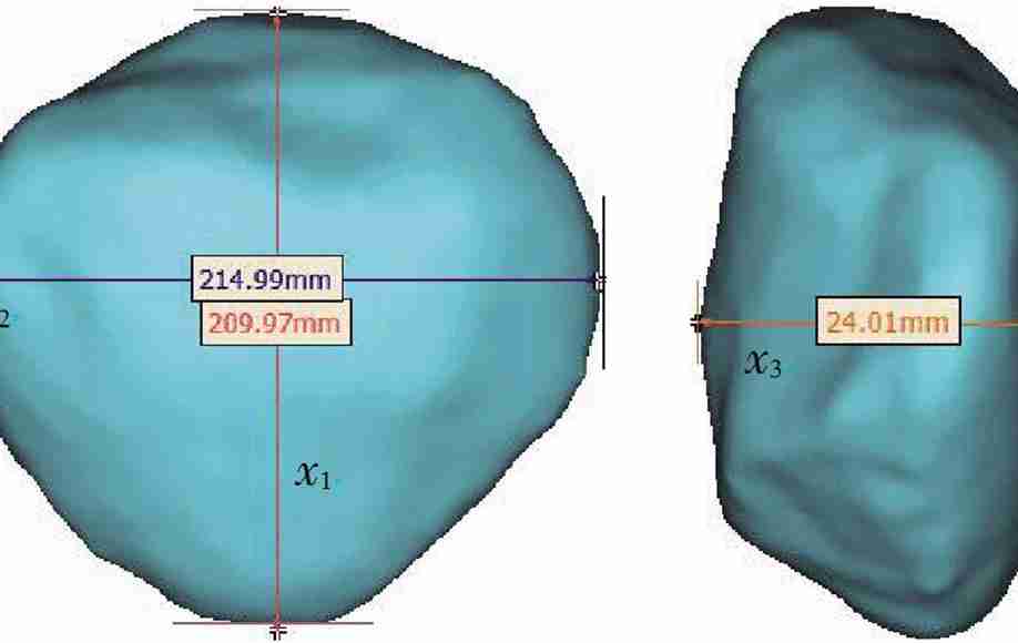
Estimation of Sex from Patella Measurements in Sichuan Han Population Based on CT-Three-Dimensional Volume Reconstruction Technique
ZHAN Meng-jun , LI Ming, LI Chun-lin , et al.
Journal of Forensic Medicine 2020, 36 (
5
): 636-641. DOI:
10.12116/j.issn.1004-5619.2020.05.006
Abstract
(
215
)
PDF(pc)
(935KB)(
785
)
Knowledge map
Save
Objective To estimate sex based on patella measurements of Sichuan Han population by computed tomography three-dimensional volume reconstruction technique, and to explore the application value of patella in sex estimation. Methods CT three-dimensional volume reconstruction images of patella of 250 individuals were collected, the four measurement indicators including patellar length, patellar width, patellar thickness, and patellar volume were measured. The t-test was used to determine measurement indicators with sex differences. Fisher discriminant analysis was used to establish the sex discriminant function and the prediction accuracy was calculated by leave-one-out cross validation. Results The sex differences of the four measurement indicators had a statistical significance (P< 0.05). The accuracy rate of the univariate discriminant function established by the patellar length was the highest (82.0%). The accuracy rates of the all indicators discriminant function and the stepwise discriminant function were 80.4% and 81.6%, respectively. Conclusion It is feasible and accurate to estimate sex of Sichuan Han population by patella measurements with CT three-dimensional volume reconstruction technique. The method may be used as an alternative for sex estimation of Sichuan Han population when other bones with higher accuracy are not available.
Related Articles
|
Metrics
|
Comments
(
0
)
Select
Sex Characteristics and Distribution of External Ear in Uygur Population of Xinjiang
MA Xin-fang , MAIMAITI Tu-di , WANG Jie-rui , et al.
Journal of Forensic Medicine 2020, 36 (
5
): 642-647. DOI:
10.12116/j.issn.1004-5619.2020.05.007
Abstract
(
3658
)
PDF(pc)
(2741KB)(
814
)
Knowledge map
Save
Objective To analyze the characteristics of the bilateral external ears of Uygur adults by directly observing the morphological characteristics of the external ears of Uygur adults and classifying each feature. The frequency distribution of the characteristics was calculated to provide reference for forensic identification. Methods The 210 cases (75 males and 135 females) of bilateral external ear photos of Uygur adults in Xinjiang that met the inclusion criteria were collected. The frequencies of the features of the external ear were recorded and distinguished between the two sexes and the different sides. The data were statistically analyzed by SPSS 21.0 statistical software. Results The shapes of the external ears of males and females were commonly oblique or rectangular (34.67% of the left external ear of males and 41.33% of the right were oblique; 30.37% of the left and right external ear of females were rectangular), while triangular ears were the rare variants and the least common. Sex and bilateral differences were observed as regards the form of the helix in the subjects. Normally rolled helix was the most common (58.67% males and 61.48% females for the left ear; 60.00% males and 72.59% females for the right ear). Wide covering scapha helix was the most rare for the male left ear and flat helix was the most rare for the female right ear. Square and free earlobes were the most common (49.33% males and 62.96% females for the left ear; 40.00% males and 54.81% females for the right ear), whereas triangular earlobes were rarely seen. Single knob tragus (40.00% males and 37.78% females for the left ear; 37.33% males and 33.33% females for the right ear) and projection type of Darwin’s tubercle (50.67% males and 40.00% females for the left ear; 48.00% males and 39.26% females for the right ear) were found to be common. Conclusion The characteristics of the bilateral external ears of male and female Uygur adults have differences, which can be used for forensic identification.
Related Articles
|
Metrics
|
Comments
(
0
)
Select
The Region of Interest in Boundary Calibration for Palatal Rugae Image of Forensic Identification
ZHANG Xiong, LI Bing, SHANGGUAN Hong, et al.
Journal of Forensic Medicine 2020, 36 (
5
): 648-653. DOI:
10.12116/j.issn.1004-5619.2020.05.008
Abstract
(
217
)
PDF(pc)
(2002KB)(
691
)
Knowledge map
Save
Objective To examine the method of least-square fitting for calibrating the palatal rugae boundary. Methods According to the distribution of the teeth, the feature points were selected; when they were fit, the boundary of the palatal rugae area was created, thereby constructing a mask. The mask was used to remove the part located outside the boundary and filter out the interference. Three samples were utilized for the experiments and analyses to come. Results To evaluate the quantitative results of the fitting curves, the correlation coefficients(r)of the samples and the relationship between the actual mean value and ideal mean value was obtained through six fitting processes. The differences between the actual mean and ideal mean were found to be significantly small(from 0.285 7 to 2.500 0)in the six fitting processes, with the range of r close to 1(from 0.989 6 to 0.999 5). Conclusion The effect of the cubic polynomial fitting method adopted in this study was stable.The proposed boundary calibration method can effectively locate the palatal rugae boundary and remove the interference area, further promoting the practice of forensic identification.
Related Articles
|
Metrics
|
Comments
(
0
)
Select
Sex Estimation of Typical Adult Vertebrae Morphology in Central China Based on CT Technique
LIU Dai-ang, YANG Li , DENG Zhen-hua , et al.
Journal of Forensic Medicine 2020, 36 (
5
): 654-659. DOI:
10.12116/j.issn.1004-5619.2020.05.009
Abstract
(
197
)
PDF(pc)
(747KB)(
778
)
Knowledge map
Save
Objective The morphological data of the second thoracic vertebra and the third lumbar vertebra were measured by computerized tomography (CT). The sex differences were analyzed and the discrimination equation was obtained. Methods The data of 274 adults (203 cases from experimental group and 69 cases from validation group) from central China were collected. Four linear data (maximum transverse length of vertebral body, maximum longitudinal length of vertebral body, maximum transverse length of vertebral foramen, maximum longitudinal length of vertebral foramen), one angle data (angle between spinous processes) and two area(vertebral foramen area, total cross-sectional area of vertebral body) data of the second thoracic vertebra and the third lumbar vertebra were collected, respectively. Then three ratios[maximum transverse length/ maximum longitudinal length of vertebral body, maximum transverse length/ maximum longitudinal length of vertebral foramen, vertebral foramen area/ (total cross-sectional area of vertebral body-vertebral foramen area)] and one angle (angle between spinous processes) were obtained. The discriminant equation was established for sexual discriminant analysis. Results The morphology of the second thoracic vertebra and the third lumbar vertebra was related with gender. Four single index discriminant formulae and eleven multi- index discriminant formulae were established. The 69 validation group samples were substituted into the formula for testing, and the maximum discriminant accuracy rate of the single-index discriminant formula was 75%. The maximum discriminant accuracy rate of multi-index discriminant formula was 83%. Conclusion It is feasible to conduct individual sex analysis by the morphological indexes of second thoracic vertebra and the third lumbar vertebra. The indexes have important application values in practice.
Related Articles
|
Metrics
|
Comments
(
0
)
 Virtual Forensic Anthropology
