法医学杂志 ›› 2022, Vol. 38 ›› Issue (1): 67-70.DOI: 10.12116/j.issn.1004-5619.2021.410607

所属专题: 水中尸体研究专题

• 论著 • 上一篇    下一篇

硅藻通过消化道淋巴系统进入实验兔体内的途径

杜宇坤1(), 刘景建2, 康晓东3, 余仲昊3, 郑冬云3, 石河3, 徐曲毅3, 任建军4, 刘超3(), 赵建3()   

  1. 1.南方医科大学法医学院,广东 广州 510515
    2.昆明医科大学法医学院,云南 昆明 650500
    3.广州市刑事科学技术研究所 法医病理学公安部重点实验室,广东 广州 510442
    4.常州大学城乡矿山研究院,江苏 常州 213164
  • 收稿日期:2020-10-27 发布日期:2022-02-25 出版日期:2022-02-28
  • 通讯作者: 刘超,赵建
  • 作者简介:赵建,男,博士研究生,主检法医师,主要从事法医病理学检验和硅藻检验:E-mail:zhaojian0721@163.com
    刘超,男,博士,主任法医师,主要从事个体识别及死因鉴定:E-mail:liuchaogzf@163.com
    杜宇坤(1997—),男,硕士研究生,主要从事法医病理学相关研究;E-mail:1105658850@qq.com
  • 基金资助:
    广州市科技计划资助项目(2019030001);公安部科技强警基础工作专项资助项目(2020GABJC38)

Pathway of Diatoms Enter Experimental Rabbits through the Lymphatic System of the Digestive Tract

Yu-kun DU1(), Jing-jian LIU2, Xiao-dong KANG3, Zhong-hao YU3, Dong-yun ZHENG3, He SHI3, Qu-yi XU3, Jian-jun REN4, Chao LIU3(), Jian ZHAO3()   
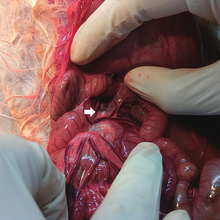
  1. 1.School of Forensic Medicine, Southern Medical University, Guangzhou 510515, China
    2.School of Forensic Medicine, Kunming Medical University, Kunming 650500, China
    3.Guangzhou Forensic Science Institute & Key Laboratory of Forensic Pathology, Ministry of Public Security, Guangzhou 510442, China
    4.Institute of Urban & Rural Mining, Changzhou University, Changzhou 213164, Jiangsu Province, China
  • Received:2020-10-27 Online:2022-02-25 Published:2022-02-28
  • Contact: Chao LIU,Jian ZHAO

摘要: 目的

研究硅藻能否通过消化道淋巴系统进入体内。

方法

将20只实验兔随机平均分为实验组和对照组,分别以20 mL珠江水样和20 mL超纯水进行灌胃,30 min后分别提取淋巴液、肺、肝和肾,对所提取样本进行硅藻检验,记录硅藻含量、大小和种类。

结果

实验组淋巴液硅藻含量高于对照组淋巴液硅藻(P<0.05)。实验组淋巴液检出冠盘藻、圆筛藻、小环藻、直链藻、菱形藻、针杆藻、桥弯藻和舟型藻,对照组检出冠盘藻、圆筛藻和小环藻。实验组淋巴液硅藻长径、短径均长于对照组(P<0.05)。实验组中,在3例肺样本、2例肝样本中检出1~2个硅藻,为冠盘藻或小环藻,肾样本中未检出硅藻;对照组中,在2例肺样本、3例肝样本中检出1~2个硅藻,为冠盘藻或圆筛藻,肾样本中未检出硅藻。

结论

硅藻可以通过淋巴液进入体内,其通过消化道淋巴系统进入体内是造成非溺死尸体组织器官内含有硅藻的原因之一。

关键词: 法医病理学, 溺死, 硅藻, 消化道, 淋巴液, 灌胃模型

Abstract: Objective

To study whether diatoms can enter the body through the lymphatic system of the digestive tract.

Methods

Twenty experimental rabbits were divided into the test group and the control group randomly, and intragastric administration was performed with 20 mL water sample from the Pearl River and 20 mL ultrapure water, respectively. After 30 min, lymph, lungs, livers and kidneys were extracted for the diatom test. The concentration, size and type of diatoms were recorded.

Results

The concentration of diatoms of the test group was higher than that of the control group (P<0.05). In the test group, Stephanodiscus, Coscinodiscus, Cyclotella, MelosiraNitzschiaSynedraCymbella, and Navicula were detected; in the control group, Stephanodiscus, Coscinodiscus and Cyclotella were detected. The long diameter and the short diameter of diatoms of the test group were higher than those of the control group (P<0.05). In the test group, 1-2 diatoms were detected in 3 lung samples and 2 liver samples, which were Stephanodiscus or Cyclotella, and no diatoms were detected in the kidney samples; in the control group, 1-2 diatoms were detected in 2 lung samples and 3 liver samples, which were Stephanodiscus or Coscinodiscus, and no diatoms were detected in the kidney samples.

Conclusion

Diatoms can enter the body through the lymphatic fluid, which is one of the reasons for the presence of diatoms in tissues and organs of non-drowning cadavers.

Key words: forensic pathology, drowning, diatom, digestive tract, lymph, gavage model

中图分类号: