Loading...

Archive

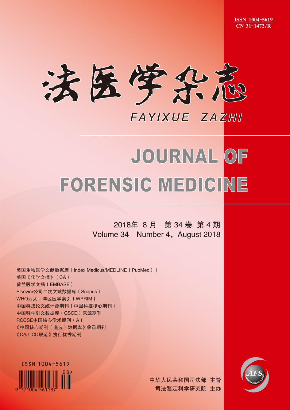
    28 August 2018, Volume 34 Issue 4 Previous Issue    Next Issue

    For Selected: Toggle Thumbnails
    Role of HMGB1 in Post-traumatic Endoplasmic Reticulum Stress in Rat Lung Tissues
    LU Jian-feng, ZHANG Qing-jie, LI Xue-hao,etal.
    2018, 34(4): 347-351.  DOI: 10.12116/j.issn.1004-5619.2018.04.001
    Abstract ( 182 )   PDF (2041KB) ( 579 )  
    Objective To explore the role of high mobility group B1 (HMGB1) protein in the post-traumatic endoplasmic reticulum stress (ERS) in rat lung tissues. Methods The rat model of acute lung injury was established by crushing the hind limbs of rats with standard weight. The first experiment was to divide rats into postural control group and crush groups (6 h, 18 h and 30 h after crushing). The second experiment was to divide rats into postural control group, 18 h crush group, HMGB1 inhibitor sodium butyrate (SB) group and 18 h crush+SB group. The protein expression changes of HMGB1 and ERS- related proteins (GRP78, caspase-12, CHOP and IRE1α) in rat lung tissues were detected with Western blotting. Meanwhile, the pathological changes of rat lungs were observed by HE stain. Results Compared with the postural control group, the expression levels of ERS-related proteins (GRP78, caspase-12, CHOP and IRE1α) and HMGB1 protein in rat lung tissues by crushing the hind limbs of rats were obviously increased. The protein levels reduced at 30 h after crushing but were still higher than those of postural control group and obvious pathological changes of acute lung injury were observed simultaneously in rats. Compared with the 18 h crush group, the expression levels of the ERS-related proteins and HMGB1 protein in rat lung tissues were attenuated in 18 h crush+SB group, and the pathological changes of rat lung injury began to alleviate. Conclusion HMGB1-ERS pathway activated by traumatic stress can lead to acute lung injury in rats.
    Related Articles | Metrics
    Estimation on Formation Time of Thrombus
    YANG Chen-teng, ZUO Min, WANG Song-jun,et al.
    2018, 34(4): 352-358.  DOI: 10.12116/j.issn.1004-5619.2018.04.002
    Abstract ( 400 )   PDF (6269KB) ( 815 )  
    Objective To observe the changes of the formation time of venous thrombus in rats, and to provide new ideas and methods for the estimation on thrombus formation time of the forensic cases died from thrombosis. Methods Totally 80 rats were randomly divided into 10 groups (0 h, 3 h, 6 h, 12 h, 1 d, 3 d, 1 week, 2 weeks, 3 weeks and 4 weeks after operation). A vein thrombosis model was established by the “narrow” method. The processes of thrombosis, organization, recanalization and the features of change on hemosiderin and calcium salt were observed by HE stain, Perls stain and Von Kossa stain. The expression changes of CD61, α-SMA and CD34 were observed by immunohistochemical staining technique. Results Platelets adhered to the exposed blood vessel intima 3 h after operation, and platelet trabeculae were formed by the repeated accumulation of platelets 1 d after operation. The thrombus organization formed through the fibroblasts from vessel wall that grew into the interior of the thrombus 3 d after operation. Endothelial cells covered the surface of thrombus and then the new blood vessels were reformed, and the vessels were reconstructed. The expression of CD61 upregulated at the stages of the thrombus formation (3 h) and thrombus reformation (4 weeks), and reached the peak 1 d after thrombus formation. The release of hemosiderin and the initial expression of α-SMA were detected 3 d later. Calcium deposit and expression of CD34 were observed 1 week later. Conclusion The hemosiderin, calcium salt, CD61, α-SMA and CD34 show time-dependent changing characteristics, which is expected to provide a reference for the estimation on thrombus formation time of the forensic cases died from thrombosis.
    Related Articles | Metrics
    Assessment of the Original Height of L1-2 after Vertebral Compression Fracture
    ZHUO Pei-pei, WANG Mao-wen, YU Xiao-ying, et al.
    2018, 34(4): 359-362.  DOI: 10.12116/j.issn.1004-5619.2018.04.003
    Abstract ( 359 )   PDF (513KB) ( 648 )  
    Objective To explore the assessment method of original height of L1-2 after vertebral compression fracture and its application value in forensic clinical practice. Methods A total of 154 normal thoracic and lumbar X-ray films were collected, and 140 cases were used as experimental group while 14 cases as validation group. The heights of anterior (Ha) and posterior (Hp) vertebral body of T12-L3 vertebrae in each X-ray image were measured. In the experimental group, the correlation analysis between HaL1 and HaT12, HpT12, HpL1, HaL2 and HpL2 was carried out, and regression equation was established via fitting. The correlation analysis between HaL2 and HaL1, HpL1, HpL2, HaL3, HpL3 was performed, and the regression equation was also established via fitting. The difference between the predicted and measured values of HaL1 and HaL2 in validation group was compared. Results In the 140 normal subjects, HaL1 (y1) was well correlated with HaT12 (x1) and HaL2 (x2), and the multiple linear regression equation was y1=2.545+0.423 x1+0.486 x2 (determining coefficient R2=0.712, P<0.05; F=169.206, P<0.05). There was no significant difference between the predicted and actual measured values of HaL1 in the validation group (P>0.05). HaL2 (y2) was well correlated with HaL1 (x3) and HaL3 (x4), and the multiple linear regression equation was y2=4.354+0.530 x3+0.349 x4 (determining coefficient R2=0.689, P<0.05; F=151.575, P<0.05). There was no significant difference between the predicted and actual measured values of HaL2 in the validation group (P>0.05). Conclusion It is more appropriate to evaluate the original height of L1 or L2 single vertebrae by comparing with the height of the anterior edge of the upper and lower adjacent vertebral bodies.
    Related Articles | Metrics
    Age Estimation and Age-related Facial Reconstruction of Xinjiang Uygur Males by Three-dimensional Human Facial Images
    PAN Si-yu, CHEN Shi-ting, TANG Kun3,et al.
    2018, 34(4): 363-369,374.  DOI: 10.12116/j.issn.1004-5619.2018.04.004
    Abstract ( 448 )   PDF (1307KB) ( 687 )  
    Objective To search age-correlated facial features and construct an age estimation model based on the three-dimensional (3D) facial images of Xinjiang Uygur males, and to structure individual face images of old age and young age. Methods Pretreatment was performed to collect 105 3D facial images of Xingjiang Uygur males aged between 17-57 years by Artec Studio software. The facial images were transferred to high-density 3D dot matrix data by FaceAnalysis software, and each image could be represented with 32 251 vertexes. Central correction of the facial images was done and all the data were aligned to a standard coordinate frame by generalized Procrustes analysis (GPA). The age estimation model was established by partial least square regression (PLSR). Furthermore, the changes of age-correlated facial features were presented on the heat map of average face, and the reconstruction of facial images at different ages was performed based on this model. Results With age, the average faces showed a series of changes including the nasolabial sulcus deepening, cheek sinking, cheekbone protruding and eye corner drooping. The Pearson correlation coefficient (r) between estimated age and chronological age was 0.71. The mean absolute deviation (MAD) of age estimation was 6.37 years. The results of age estimation in >30-40 years group showed a best accuracy (MAD=4.27 years), and the deviations increased with age after 40 years. The composite facial images represented a significant result with age on facial morphological features and aging. Conclusion The results of this study reveal the age-correlated facial features and aging markers in Uygur population, which help to construct a reliable age estimation model.
    References | Related Articles | Metrics
    Formula Derivation for the Probability Distribution of IBS Score in Unrelated Individual Pairs
    ZHAO Huan-dong, ZHAO Shu-min, CHEN Yu-xiang, et al.
    2018, 34(4): 370-374.  DOI: 10.12116/j.issn.1004-5619.2018.04.005
    Abstract ( 342 )   PDF (629KB) ( 488 )  
    Objective To derive the probability equation given by STR allele frequencies of identity by state (IBS) score shared by unrelated individual pairs. Methods By comparing the STR genotypes of two unrelated individuals, three mutually exclusive combinations could be obtained: (1) sharing 2 identical alleles, a2=1, otherwise a2=0; (2) sharing 1 identical allele, a1=1, otherwise a1=0; (3) sharing 0 identical allele, a0=1, otherwise a0=0. And the IBS score of the one STR locus in this unrelated individual pair could be given by the formula: ibs=2a2+a1. The probability of a2=1 (p2), a1=1 (p1) and a0=1 (p0) were derived and expressed in powers of the allele frequencies. Subsequently, for a genotyping system including n independent STR loci, the characteristics of binomial distribution of IBS score shared by a pair of unrelated individuals could be given by p2l and p1l (l=1, 2, …, n). Results All the general equations of p2, p1 and p0 were derived from the basic conceptions of a2, a1 and a0, respectively. Given fi (i=1, 2, …, m) as the ith allele frequency of a STR locus, the general equations of p2, p1 and p0 could be respectively expressed in powers of fi: p2=2(■fi2)2-■fi4, p1=4■fi2-4(■fi2)2-4■fi3+4■fi4 and p0=1-4■fi2+2(■fi2)2+4■fi3-3■fi4. The sum of p2, p1 and p0 must be equal to 1. Then, the binomial distribution of IBS score shared by unrelated individual pairs genotyped with n independently STR loci could be written by: IBS~B(2n, π), and the general probability, π, could be given by the formula: π=■■p2l+■■p1l. Conclusion In the biological full sibling identification, the probability of null hypothesis corresponding to any specific IBS score can be directly calculated by the general equations presented in this study, which is the basement of the evidence explanation.
    References | Related Articles | Metrics
    Rapid Determination of Cocaine and Its Metabolite Benzoylecgonine in Hair by LC-MS/MS
    PAN Mei-ru, QIANG Huo-sheng, SHEN Bao-hua,et al.
    2018, 34(4): 375-378,383.  DOI: 10.12116/j.issn.1004-5619.2018.04.006
    Abstract ( 374 )   PDF (731KB) ( 766 )  
    Objective To establish a rapid determination method with LC-MS/MS for cocaine and its metabolite benzoylecgonine in hair. Methods Deuterated internal standards (cocaine-D3 and benzoylecgonine-D8) were added to the decontaminated hair. After the extraction by ultrasonication with methanol, the compounds were separated by the Restek Allure PFP propyl column, and cocaine and benzoylecgonine were simultaneously analysed in multiple reaction monitoring mode. Results The cocaine and benzoylecgonine in hair showed a good linearity in the range of mass fraction between 0.02 and 10.00 ng/mg with the limits of detection of 0.01 ng/mg. Conclusion The developed method is simple and rapid with a good selectivity, which is suitable for the determination of cocaine and its metabolite benzoylecgonine in hair.
    References | Related Articles | Metrics
    Determination of Endosulfan Concentrations in Biological Samples by GC-MS/MS
    ZHANG Fan, QIAO Jun-yuan, YU Ming-jun,et al .
    2018, 34(4): 379-383.  DOI: 10.12116/j.issn.1004-5619.2018.04.007
    Abstract ( 275 )   PDF (720KB) ( 519 )  
    Objective To establish an analytical method of the endosulfan concentrations (α-endosulfan and β-endosulfan) in biological samples by GC-MS/MS. To observe the distribution of endosulfan in aquatic animals and provide experimental evidence for forensic identification of relevant cases. Methods Acetonitrile was added to the blood and muscle samples for precipitating the protein. The endosulfan concentrations were determined by GC-MS/MS in multiple reaction monitoring mode. Qualitative analysis was performed according to the retention time and ion rate, and quantitative analysis was performed by external standard working curve method. Results In blood samples, the calibration curves of α-endosulfan and β-endosulfan ranging from 0.062 5 to 10 μg/mL had good linear relationship, the correlation coefficients (r) of which were >0.99. The limits of detection (LOD) were 1 ng/mL and 2 ng/mL and the limits of quantification (LOQ) were 4 ng/mL and 8 ng/mL, respectively. In muscle samples, the calibration curves of α-endosulfan and β-endosulfan ranging from 0.062 5 to 10 μg/g, the r of which were >0.98. The LOD were 1 ng/g and 4 ng/g and the LOQ were 4 ng/g and 16 ng/g, respectively. The accuracy of α-endosulfan and β-endosulfan was 90.76%-108.91% both in blood and muscle samples, the interday and intraday precision were 2.35%-8.71% and 5.44%-10.29%, respectively. In poisoning cases, endosulfan were detected in all parts of fish and crab and the content difference was statistically significant. Conclusion The endosulfan detection method based on GC-MS/MS established in the present study is rapid, sensitive and accurate, which can be applied to the endosulfan detection in traces biological samples. The distribution of endosulfan in fish and crab was different, which can provide evidence to the sample collection and analysis for toxicological analysis in relevant forensic identification.
    Related Articles | Metrics
    Forensic Pathology Analysis of 363 Sudden Death Cases in Yunnan Province
    SUN Zhong-chun, YANG Qi-kun, JIA Peng-lin,et al.
    2018, 34(4): 384-388.  DOI: 10.12116/j.issn.1004-5619.2018.04.008
    Abstract ( 318 )   PDF (686KB) ( 894 )  
    Objective To study the epidemiological and pathological features of sudden death (SD) in Yunnan Province and to provide scientific evidence for prevention and forensic identification of sudden death. Methods Totally 363 SD cases were collected from the autopsies between 2009 and 2017 in the Forensic Centre of Kunming Medical University. The related factors such as etiology, age, inducing factor, time interval between the onset of disease and death, morbidity season and pathological change were retrospectively analysed. Results The incidence of SD in males was significantly higher than that of females. The peak age was ≥35-55 years. The mortality rate was relatively high within 6 h after the onset of disease. The season order with descending number of deaths was spring, summer, winter and autumn. The top ten causes of SD were coronary heart disease, sudden unexplained death (SUD), cerebral hemorrhage, acute hemorrhagic necrotic pancreatitis, aortic dissection rupture, cardiomyopathy, pneumonia, pulmonary thromboembolism, amniotic fluid embolism and allergy. Exercise, infusion, surgery, medication and minor injury were the most common predisposing factors of sudden coronary death. Consciousness disorder or coma, chest pain or chest tightness, and abdominal pain were the most common premortem symptoms of sudden coronary death. Conclusion The SD is more common in middle-aged males, which is the key population for the prevention of SD. For the forensic identification and prevention of SD, the attention on SUD should be paid.
    Related Articles | Metrics
    Forensic Analysis on 52 Medical Malpractice Cases of Cardiac Death
    LU Jia, ZHANG Yun-lou, LUO Lin
    2018, 34(4): 389-391,395.  DOI: 10.12116/j.issn.1004-5619.2018.04.009
    Abstract ( 272 )   PDF (631KB) ( 531 )  
    Objective To synthetically analyse the medical malpractice cases of cardiac death in forensic identification, and to explore the generality and characteristic of cause of death, medical malpractice and assessment of participation degree. Methods Totally 52 medical malpractice cases of cardiac death examined in the Xiaoshan Branch Office, Hangzhou Minghao Forensic Judical Appraisal Institute, from January 2015 to April 2018 were collected. The general information of cases, medical institutions and situations of hospital stay, cause of death, medical malpractice and assessment of participation degree were retrospectively analysed. Results In 52 cases, the male to female ratio was 2.25:1, and most subjects aged >50-60. Cardiac death caused by hypertensive heart disease or coronary heart disease was most common (67.3%), followed by viral myocarditis and cardiomyopathy (13.5%). There were 24 cases involved surgery, and the survival time after surgery was from 1 h to 118 d with a 7 d medium value. There were 63 medical institutes involved in these medical malpractices. Medical malpractice presented in most hospitals more or less, and the participation degree was >20%-30%. Conclusion Forensic appraisal contributes to determine causes of death, which not only provides scientific evidence for medical malpractice identification, but also improves diagnosis and treatment levels of medical institutions.
    References | Related Articles | Metrics
    Retrospective Analysis of 24 Cases of Forensic Medical Identification on Traumatic Tympanic Membrane Perforations#br#
    CHEN Fang, YANG Xiao-ping, LIU Xia,et al.
    2018, 34(4): 392-395.  DOI: 10.12116/j.issn.1004-5619.2018.04.010
    Abstract ( 237 )   PDF (1492KB) ( 569 )  
    Objective To study the case characteristics of forensic medical identification of traumatic tympanic membrane perforations, and to discuss the key points of forensic medical identification and evaluations methods for tympanic membrane perforations. Methods Twenty-four cases of traumatic tympanic membrane perforations accepted by the Academy of Forensic Science during 2017 were retrospectively analysed. The data of perforation size, form, predilection site, healing time and healing mode were evaluated. Results For the traumatic tympanic membrane perforations, the study showed that the small size of perforation (<1/2 quadrant) with irregular shape was common. The location of perforations was almost on the anterior and inferior quadrant, and centripetal migration healing was common. The healing rate within 6 weeks was up to 90%. Conclusion In the identification cases of traumatic tympanic membrane perforations, the key is to determine whether it is traumatic and whether it will heal spontaneously within 6 weeks. It is suggested to check the tympanic membrane weekly by an otic endoscope combined with acoustic impedance measurement at the sixth week, which can improve the accuracy, objectivity and scientificity of the identification.
    Related Articles | Metrics
    Detection and Analysis of 12 Suspected Amelogenin Allelic Loss Cases
    BI Jie, CHANG Jing-jing, YU Chun-ying
    2018, 34(4): 396-400.  DOI: 10.12116/j.issn.1004-5619.2018.04.011
    Abstract ( 247 )   PDF (925KB) ( 605 )  
    Objective To observe and analyse the Amelogenin allelic loss in parent-child identification cases, and to explore the type and mechanism of Amelogenin allelic loss as well as its influence on gender identification and solutions. Methods After the detection by SiFaSTRTM 23plex DNA identification system, samples had the characteristics of the peak area of Amelogenin X was the same as the one of adjacent heterozygote or lower than one half of adjacent homozygote in females while Amelogenin X loss was observed in males were selected. X chromosome STR (X-STR) typing and Amelogenin X sequencing were performed. The samples with Amelogenin Y loss in males were confirmed by the detection of Y chromosome STR typing and sex-determining region of Y (SRY). The type and rate of Amelogenin allelic loss were confirmed and calculated, and the mechanism and influence of this variation were also analysed. Results Amelogenin X allelic loss was observed in one male sample, the mutation in primer-binding region was confirmed by sequencing. The suspected Amelogenin X allelic loss was observed in four female samples, but the mutation in primer-binding region was confirmed by sequencing in only one sample. Amelogenin Y allelic loss was observed in seven male samples, SRY positive cases was detected in five of them, and two were SRY negative. Y-STR type was detected in four cases of the five SRY positive cases, which was not detected in the two SRY negative cases. The rate of Amelogenin allelic loss was about 0.029%. Conclusion Amelogenin X allelic loss does not affect the gender identification, but Amelogenin Y allelic loss may cause wrong gender identification. Thus, Y-STR or SRY should be detected for gender confirmation. When Y-STR genotypes are not detected in a “male” whose SRY detection is also negative, then the chromosome karyotype analysis and sex differentiation related genes test should be taken to further confirm the gender.
    Related Articles | Metrics
    Comparison of Single Piece of Dandruff DNA Extraction under Microscope and EZ-tape Method
    BAI Xiao-gang, JIAN Hui, WANG Hui, et al.
    2018, 34(4): 401-404.  DOI: 10.12116/j.issn.1004-5619.2018.04.012
    Abstract ( 206 )   PDF (1161KB) ( 558 )  
    Objective To collect single piece of dandruff with microscopes to improve the regular EZ-tape method for DNA extraction and genotyping, increase the utilization of samples, reduce the miss rate as well as the proportion of genotyping results of mixed stains. Methods The insides of the hats worn by two volunteers were stuck by EZ-tape and scotch tape respectively. DNA on EZ-tape was directly extracted using traditional method. Single piece of dandruff was collected from the scotch tapes under microscope. The two kinds of methods were both performed under continuous oscillation and standing digestion, respectively. DNA was extracted through Chelex-100 method, and STR genotypes were obtained after amplification and electrophoresis. The results of STR genotypes obtained by EZ-tape method and single piece of dandruff analytical method were compared. Results Miss detections happened in 11 samples (45.8%) by EZ-tape method and only single-source typing results were obtained. Ten samples (41.7%) showed the genotype results of mixed stain and six of which showed allele insertions and deletions. The genotype results were obtained successfully using the single piece of dandruff analytical method, and two samples showed mixed stain genotype. The number of exact typing processed by oscillation was higher than that by standing digestion (P<0.05). Conclusion The oscillation during the DNA extraction process is in favour of the DNA releasing. Single piece of dandruff analytical method can be used to obtain single-source STR genotype with high successful ratio and low miss rate. This method can be a collection method of special samples such as dandruff in forensic practice.
    Related Articles | Metrics
    Application of Multiple Kits in Special Parentage Testing Cases
    GAO Hong-mei, WANG Chang, ZHANG Shan-shan,et al.
    2018, 34(4): 405-410,416.  DOI: 10.12116/j.issn.1004-5619.2018.04.013
    Abstract ( 237 )   PDF (715KB) ( 579 )  
    Objective To analyse the genetic polymorphism of 21 autosome STR loci in Han population of Shandong Province and the cases with loci mutation or allelic loss typed by Goldeneye?誖DNA identification system 25A. Methods Totally 40 autosome STR loci types of 273 unrelated individuals in Han population of Shandong Province were typed by Goldeneye?誖DNA identification system 25A and 22NC, and the genetic polymorphism of 21 STR loci in those was analysed. Meanwhile, six cases with loci mutation were analysed by adding the tests with Goldeneye?誖DNA identification system 22NC, 20Y and 17X. Another three cases with allelic loss were tested by AmpF?詛STR?誖Identifiler?誖Plus PCR and analysed by gene sequencing. Results The genetic parameters of 21 autosome STR loci in Han population of Shandong Province were obtained. When STR loci were added up to 40, five of those with loci mutation met the identification requirements, and the results of X-STR or Y-STR types were consistent with that of STR loci. There was another duo case with one suspected loci mutation, biological source of six STR loci genotypes could not be found in the genotypes of supposed father. The Y-STR genotype of two individuals was identical that indicated both of them came from same paternal line. However, the fatherhood was excluded according to the autosome STR loci system. For two cases with allelic loss on D18S51, base mutation or loss were found in the primer binding domain of mother and child by gene sequencing. Another mother-child case with allelic loss on D13S317 was certified by AmpF?詛STR?誖Identifiler?誖Plus PCR kit. Conclusion The 21 autosome STR loci in Han population of Shandong Province have high polymorphism, which can be used in routine cases of paternity identification. For some duo cases with loci mutation, Goldeneye?誖DNA identification system 25A cannot satisfy the identification requirements, thus more autosome STR loci should be added properly. For the cases with allelic loss, the problem can be resolved by gene sequencing or using different merchant kits.
    References | Related Articles | Metrics
    Genetic Polymorphisms and Mutations of 30 Y-STR Loci in Chinese Han Population
    WU Wei-wei, SU Yan-jia, MEI Xing-lin,et al.
    2018, 34(4): 411-416.  DOI: 10.12116/j.issn.1004-5619.2018.04.014
    Abstract ( 202 )   PDF (559KB) ( 742 )  
    Objective To investigate the genetic polymorphisms and mutations of 30 Y-STR loci in Chinese Han males and to evaluate its forensic application. Methods The DNA extracted from blood samples of 1 005 unrelated males and 1 008 father-son pairs (1 949 individuals in all) in Chinese Han population were typed using developed 30 Y-STR loci identification system. The parameters of population genetics and the mutation rates of each locus were analysed statistically. Results A total of 983 haplotypes were found in 1 005 unrelated males from Chinese Han population, of which 963 were unique. The overall haplotype diversity (HD) and discrimination capacity (DC) were 0.999 955 and 0.978 109, respectively. Totally 340 alleles were detected on 30 Y-STR loci, the value of gene diversity (GD) ranged from 0.410 3 to 0.952 3. The GD values of 24 out of the 30 loci were over 0.6. There were 30 269 allele transfers in 1 008 father-son pairs, one mutation in 68 father-son pairs, and the mutation of three father-son pairs occurred at two loci. On 26 Y-STR loci, 74 mutations were detected in 71 father-son pairs. The average mutation rates were 2.4×10⁻³ (95% CI: 1.9×10⁻³-3.1×10⁻³). Seventy-three mutation events were one-step mutation (98.6%), 1 mutation event was two-step mutation (1.4%). Conclusion The multiplex PCR system with 30 Y-STR loci has high genetic polymorphism and low mutation rates in Chinese Han males. Therefore, the system shows important values in Y-STR database construction and population genetic research.
    Related Articles | Metrics
    Extraction of DNA from Sperm Cells in Mixed Stain by Nylon Membrane Bushing Separation Technique
    MA Jun, TONG Qi, GAO Liang-bi,et al.
    2018, 34(4): 417-419,427.  DOI: 10.12116/j.issn.1004-5619.2018.04.015
    Abstract ( 264 )   PDF (518KB) ( 586 )  
    Objective To establish a novel method for the separation of sperm cells in mixed stain, and to evaluate its application value. Methods Totally 40 mixed stain samples were collected from sexual assault cases. Sperm cells were separated by the conventional differential lysis method and the nylon membrane bushing separation technique, respectively. The DNA of sperm cells was extracted with the silicon membrane kit (Forensic DNA Extraction Kit for Soft Tissues). The PCR amplification was performed using AmpF?詛STR?誖Identifiler?誖Plus kit, and the products were electrophoresed by 3500xL genetic analyser. The results of two separation methods were then compared. Results Complete and single-source male STR genotypes could be obtained from all the 40 mixed stain samples except three samples with minimal residual of female DNA by the nylon membrane bushing separation technique. The STR genotypes of sperm cells could not be detected in 25 samples, which were obtained in 15 samples (seven were of incomplete male STR genotypes, six with residual of female DNA, two were complete and single-source STR genotypes of sperm cells). Conclusion The nylon membrane bushing separation technique developed in present study can be used in the separation of sperm cells in mixed stain, especially for the extraction of a small amount of sperm from a large quantity of female cells, which is inexpensive, rapid and simple.
    References | Related Articles | Metrics
    Research Progress on InDel Genetic Marker in Forensic Science
    SHENG Xiang, BAO Yun, ZHANG Jia-shuo,et al.
    2018, 34(4): 420-427.  DOI: 10.12116/j.issn.1004-5619.2018.04.016
    Abstract ( 736 )   PDF (816KB) ( 1228 )  
    Genetic markers in forensic DNA typing experienced the variable number of tandem repeats (VNTR) sequences and the short tandem repeats (STR) sequences. With the emerge of sequencing technology, the third generation of genetic markers were found out, which usually have two alleles including single nucleotide polymorphism (SNP) and insertion/deletion (InDel), also known as biallelic genetic markers. Because of the insertions or deletions of DNA fragments, InDel genetic marker reveals DNA fragment length polymorphism and widely distributes across the whole genome. InDel genetic marker is numerous and has the characteristics of STR and SNP genetic markers, which has been applied in the fields of genetics and anthropology. This review focuses on the research progress of InDel genetic marker in forensic science, aiming to review and summarize the main research findings in recent years and provide clues for future researches.
    Related Articles | Metrics
     
    2018, 34(4): 432-434,437.  DOI: 10.12116/j.issn.1004-5619.2018.04.019
    Abstract ( 188 )   PDF (652KB) ( 532 )  
     
    References | Related Articles | Metrics