Journal of Forensic Medicine ›› 2025, Vol. 41 ›› Issue (3): 201-207.DOI: 10.12116/j.issn.1004-5619.2024.441004
• Original Articles • Next Articles
Qian WANG1,2( ), Song-min YANG3, Juan-juan WU1,2, Yu ZHANG1,2, Xiang-meng WANG1,2, Gang CHEN1,2, Peng-fei JIANG1()
Received:2024-10-09
Online:2025-09-15
Published:2025-06-25
Contact:
Peng-fei JIANG
CLC Number:
Qian WANG, Song-min YANG, Juan-juan WU, Yu ZHANG, Xiang-meng WANG, Gang CHEN, Peng-fei JIANG. Temporal Expression of NETosis Marker CitH3 in Deep Vein Thrombosis in Mice[J]. Journal of Forensic Medicine, 2025, 41(3): 201-207.
Add to citation manager EndNote|Ris|BibTeX
URL: http://www.fyxzz.cn/EN/10.12116/j.issn.1004-5619.2024.441004
| 组别 | CitH3阳性细胞数量 | 中性粒细胞数量 | C/N |
|---|---|---|---|
| 建模后0 h | 0.4 | 8.6 | 3.5 |
| 建模后1 d | 26.0 | 96.4 | 26.9 |
| 建模后3 d | 40.4 | 77.8 | 52.0 |
| 建模后5 d | 48.4 | 56.8 | 85.6 |
| 建模后7 d | 25.2 | 47.8 | 53.1 |
| 建模后10 d | 3.4 | 43.4 | 7.7 |
| 建模后14 d | 1.2 | 35.0 | 3.2 |
| 建模后21 d | 0.6 | 20.2 | 2.8 |
Tab. 1 The numbers of CitH3-positive cells andneutrophils, as well as their ratios in each group
| 组别 | CitH3阳性细胞数量 | 中性粒细胞数量 | C/N |
|---|---|---|---|
| 建模后0 h | 0.4 | 8.6 | 3.5 |
| 建模后1 d | 26.0 | 96.4 | 26.9 |
| 建模后3 d | 40.4 | 77.8 | 52.0 |
| 建模后5 d | 48.4 | 56.8 | 85.6 |
| 建模后7 d | 25.2 | 47.8 | 53.1 |
| 建模后10 d | 3.4 | 43.4 | 7.7 |
| 建模后14 d | 1.2 | 35.0 | 3.2 |
| 建模后21 d | 0.6 | 20.2 | 2.8 |
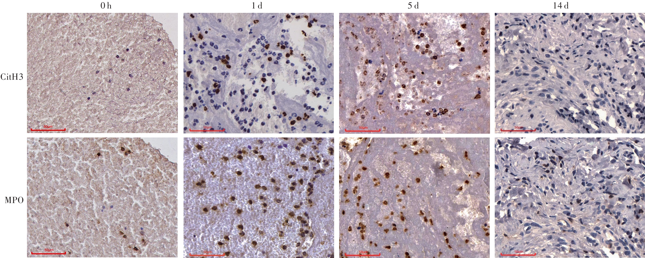
Fig. 1 Immunohistochemical staining results of CitH3 and MPO during the development of IVC thrombosis in mice
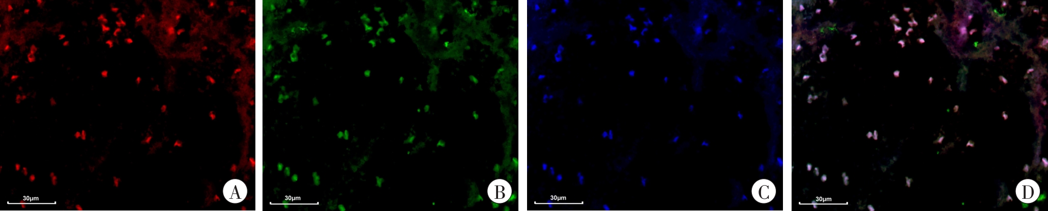
Fig. 2 Double immunofluorescence staining results of CitH3 and MPO in thrombus at 3 d after modeling
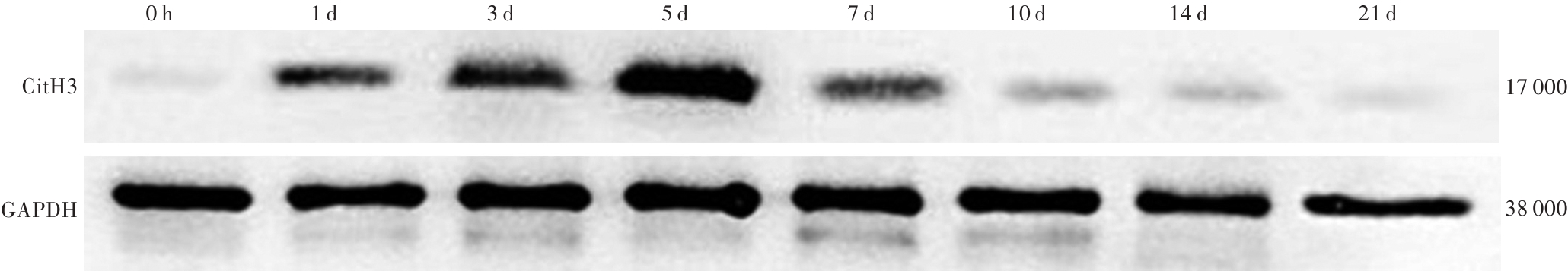
Fig. 3 Western blotting analysis of CitH3 protein expression at different time points after modeling
| 组别 | CitH3蛋白 |
|---|---|
| 建模后0 h | 0.03 |
| 建模后1 d | 0.53 |
| 建模后3 d | 0.74 |
| 建模后5 d | 0.98 |
| 建模后7 d | 0.47 |
| 建模后10 d | 0.15 |
| 建模后14 d | 0.09 |
| 建模后21 d | 0.04 |
Tab. 2 Relative expression levels ofCitH3 protein in each group
| 组别 | CitH3蛋白 |
|---|---|
| 建模后0 h | 0.03 |
| 建模后1 d | 0.53 |
| 建模后3 d | 0.74 |
| 建模后5 d | 0.98 |
| 建模后7 d | 0.47 |
| 建模后10 d | 0.15 |
| 建模后14 d | 0.09 |
| 建模后21 d | 0.04 |
| [1] | KHAN F, TRITSCHLER T, KAHN S R, et al. Venous thromboembolism[J]. Lancet,2021,398(10294):64-77. doi:10.1016/S0140-6736(20)32658-1 . |
| [2] | LUTSEY P L, ZAKAI N A. Epidemiology and prevention of venous thromboembolism[J]. Nat Rev Cardiol,2023,20(4):248-262. doi:10.1038/s41569-022-00787-6 . |
| [3] | FANOLA C L, NORBY F L, SHAH A M, et al. Incident heart failure and long-term risk for venous thromboembolism[J]. J Am Coll Cardiol,2020,75(2):148-158. doi:10.1016/j.jacc.2019.10.058 . |
| [4] | CHOPARD R, ALBERTSEN I E, PIAZZA G. Diagnosis and treatment of lower extremity venous thromboembolism: A review[J]. JAMA,2020,324(17):1765-1776. doi:10.1001/jama.2020.17272 . |
| [5] | HUANG J J, ZHUO J Y, WANG Q, et al. The time-dependent expression of FPR2 and ANXA1 in murine deep vein thrombosis model and its relation to thrombus age[J]. Forensic Sci Med Pathol,2024,20(4):1155-1165. doi:10.1007/s12024-024-00818-3 . |
| [6] | 吴娟娟,黄俊杰,张煜,等. IL-10、TGF-β1在小鼠深静脉血栓中的时序性变化[J].法医学杂志,2024,40(2):179-185. doi:10.12116/j.issn.1004-5619.2023.430506 . |
| WU J J, HUANG J J, ZHANG Y, et al. Time-dependent sequential changes of IL-10 and TGF-β1 in mice with deep vein thrombosis[J]. Fayixue Zazhi,2024,40(2):179-185. | |
| [7] | NOSAKA M, ISHIDA Y, KIMURA A, et al. Time-dependent appearance of intrathrombus neutrophils and macrophages in a stasis-induced deep vein thrombosis model and its application to thrombus age determination[J]. Int J Legal Med,2009,123(3):235-240. doi:10.1007/s00414-009-0324-0 . |
| [8] | JAILLON S, PONZETTA A, DI MITRI D, et al. Neutrophil diversity and plasticity in tumour progression and therapy[J]. Nat Rev Cancer,2020,20(9):485-503. doi:10.1038/s41568-020-0281-y . |
| [9] | CASTANHEIRA F V S, KUBES P. Neutrophils and NETs in modulating acute and chronic inflammation[J]. Blood,2019,133(20):2178-2185. doi:10.1182/blood-2018-11-844530 . |
| [10] | BRILL A, FUCHS T A, CHAUHAN A K, et al. von Willebrand factor-mediated platelet adhesion is critical for deep vein thrombosis in mouse models[J]. Blood,2011,117(4):1400-1407. doi:10.1182/blood-2010-05-287623 . |
| [11] | ETULAIN J, MARTINOD K, WONG S L, et al. P-selectin promotes neutrophil extracellular trap formation in mice[J]. Blood,2015,126(2):242-246. doi:10.1182/blood-2015-01-624023 . |
| [12] | BRINKMANN V, REICHARD U, GOOSMANN C, et al. Neutrophil extracellular traps kill bacteria[J]. Science,2004,303(5663):1532-1535. doi:10.1126/science.1092385 . |
| [13] | BRILL A, FUCHS T A, SAVCHENKO A S, et al. Neutrophil extracellular traps promote deep vein thrombosis in mice[J]. J Thromb Haemost,2012,10(1):136-144. doi:10.1111/j.1538-7836.2011.04544.x . |
| [14] | DYER M R, CHEN Q W, HALDEMAN S, et al. Deep vein thrombosis in mice is regulated by platelet HMGB1 through release of neutrophil-extracellular traps and DNA[J]. Sci Rep,2018,8(1):2068. doi:10.1038/s41598-018-20479-x . |
| [15] | THÅLIN C, LUNDSTRÖM S, SEIGNEZ C, et al. Citrullinated histone H3 as a novel prognostic blood marker in patients with advanced cancer[J]. PLoS One,2018,13(1):e0191231. doi:10.1371/jour nal.pone.0191231 . |
| [16] | THÅLIN C, HISADA Y, LUNDSTRÖM S, et al. Neutrophil extracellular traps: Villains and targets in arterial, venous, and cancer-associated thrombosis[J]. Arterioscler Thromb Vasc Biol,2019,39(9):1724-1738. doi:10.1161/ATVBAHA.119.312463 . |
| [17] | 尧梦婷,方储存,王子龙,等. 中性粒细胞胞外诱捕网在深静脉血栓形成机制中的研究进展[J].赣南医学院学报,2024,44(4):343-348. doi:10.3969/j.issn.1001-5779.2024.04.003 . |
| YAO M T, FANG C C, WANG Z L, et al. Research progress of neutrophil extracellular traps in deep vein thrombosis mechanism[J]. Gannan Yixue-yuan Xuebao,2024,44(4):343-348. | |
| [18] | SARAVANAN R, CHOONG Y K, LIM C H, et al. Cell-free DNA promotes thrombin autolysis and ge-neration of thrombin-derived C-terminal fragments[J]. Front Immunol,2021,12:593020. doi:10.3389/fimmu.2021.593020 . |
| [19] | MASUDA S, NAKAZAWA D, SHIDA H, et al. NETosis markers: Quest for specific, objective, and quantitative markers[J]. Clin Chim Acta,2016,459:89-93. doi:10.1016/j.cca.2016.05.029 . |
| [20] | HAKKIM A, FUCHS T A, MARTINEZ N E, et al. Activation of the Raf-MEK-ERK pathway is required for neutrophil extracellular trap formation[J]. Nat Chem Biol,2011,7(2):75-77. doi:10.1038/nchembio.496 . |
| [21] | LI P X, LI M, LINDBERG M R, et al. PAD4 is essential for antibacterial innate immunity mediated by neutrophil extracellular traps[J]. J Exp Med,2010,207(9):1853-1862. doi:10.1084/jem.20100239 . |
| [22] | MAURACHER L M, POSCH F, MARTINOD K, et al. Citrullinated histone H3, a biomarker of neutrophil extracellular trap formation, predicts the risk of venous thromboembolism in cancer patients[J]. J Thromb Haemost,2018,16(3):508-518. doi:10.1111/ jth.13951 . |
| [23] | BOETTCHER M, ESSER M, TRAH J, et al. Markers of neutrophil activation and extracellular traps formation are predictive of appendicitis in mice and humans: A pilot study[J]. Sci Rep,2020,10(1):18240. doi:10.1038/s41598-020-74370-9 . |
| [24] | NOMURA K, MIYASHITA T, YAMAMOTO Y, et al. Citrullinated histone H3: Early biomarker of neutrophil extracellular traps in septic liver damage[J]. J Surg Res,2019,234:132-138. doi:10.1016/j.jss.2018.08.014 . |
| [25] | EL-SAYED O M, DEWYER N A, LUKE C E, et al. Intact Toll-like receptor 9 signaling in neutrophils modulates normal thrombogenesis in mice[J]. J Vasc Surg,2016,64(5):1450-1458.e1. doi:10.1016/ |
| j.jvs.2015.08.070. | |
| [26] | NOSAKA M, ISHIDA Y, KUNINAKA Y, et al. Relationship between intrathrombotic appearance of HSP27 and HSP70 and thrombus ages in a murine model of deep vein thrombosis[J]. Sci Rep,2023,13(1):22416. doi:10.1038/s41598-023-48987-5 . |
| [27] | NOSAKA M, ISHIDA Y, KUNINAKA Y, et al. Intrathrombotic appearances of AQP-1 and AQP-3 in relation to thrombus age in murine deep vein thrombosis model[J]. Int J Legal Med,2021,135(2):547-553. doi:10.1007/s00414-020-02482-y . |
| [28] | IRNIGER W. Histologic age determination of thrombi and emboli[J]. Virchows Arch Pathol Anat Physiol Klin Med,1963,336(3):220-237. doi:10.1007/BF00957911 . |
| [29] | FINESCHI V, TURILLAZZI E, NERI M, et al. Histological age determination of venous thrombosis: A neglected forensic task in fatal pulmonary thrombo-embolism[J]. Forensic Sci Int,2009,186(1/2/3):22-28. doi:10.1016/j.forsciint.2009.01.006 . |
| [30] | NICKLAS J M, GORDON A E, HENKE P K. Resolution of deep venous thrombosis: Proposed immune paradigms[J]. Int J Mol Sci,2020,21(6):2080. doi:10.3390/ijms21062080 . |
| [31] | NAJEM M Y, COUTURAUD F, LEMARIÉ C A. Cytokine and chemokine regulation of venous thromboembolism[J]. J Thromb Haemost,2020,18(5):1009-1019. doi:10.1111/jth.14759 . |
| [32] | MUKHOPADHYAY S, JOHNSON T A, DURU N, et al. Fibrinolysis and inflammation in venous thrombus resolution[J]. Front Immunol,2019,10:1348. doi:10.3389/fimmu.2019.01348 . |
| [33] | CAMPOS J, BRILL A. By word of mouse: Using animal models in venous thrombosis research[J]. Platelets,2020,31(4):447-454. doi:10.1080/09537104.2019.1678117 . |
| [34] | 张茜,谷田,刘哲宇,等. 小鼠深静脉血栓实验模型的制备[J].江苏大学学报(医学版),2022,32(5):398-402. doi:10.13312/j.issn.1671-7783.y220140 . |
| ZHANG X, GU T, LIU Z Y, et al. Preparation of experimental animal model of mouse deep vein thrombosis[J]. Jiangsu Daxue Xuebao (Medicine edition),2022,32(5):398-402. |
| [1] | Xiao-feng ZHANG, Qin SU, Xiao-hui CHEN, Wei-bin WU, Dong-yun ZHENG, Jian ZHAO, Ling CHEN, Qu-yi XU, Chao LIU. Comparison of Three Drowning‑related Plankton Testing Methods in Drowning Diagnosis [J]. Journal of Forensic Medicine, 2025, 41(3): 244-251. |
| [2] | Xuan-long CHEN, Qiang YUAN, Yong SUN, Die ZHANG, Jian-bin FU, Li-liang LI. Forensic Research Progress on Bongkrekic Acid Poisoning [J]. Journal of Forensic Medicine, 2025, 41(2): 111-119. |
| [3] | Shuai ZHANG, Hong-fei XU, Zhi-xiang ZHANG, Ying WANG, Shao-hua ZHU. Research on Doxorubicin-Induced Cardiotoxicity Mechanism and Its Forensic Application [J]. Journal of Forensic Medicine, 2025, 41(2): 120-126. |
| [4] | Yu-meng ZUO, Wei HAN, Jian-bo ZHANG, Tao LI. Molecular Mechanisms and Toxic Effects of Ketamine [J]. Journal of Forensic Medicine, 2025, 41(2): 127-135. |
| [5] | Rong-shuai WANG, Si-zhe HUANG, Yun-yun WANG, Yan-fei DENG, Zi-jiao DING, Jie ZHANG, Yong LIU, Liang REN, Liang LIU. The Mechanism of Calcium Handling Proteins and NF-κB in Calcium Dyshomeostasis of Cardiomyocytes Caused by Acute MDMA Exposure [J]. Journal of Forensic Medicine, 2025, 41(2): 144-151. |
| [6] | Hao-wei WANG, Xiao-xing ZHANG, Gen-meng YANG, Shang-wen WANG, Xiao-feng ZENG. The Role of Ferroptosis in Hepatocyte Injury Induced by α-Amanitin [J]. Journal of Forensic Medicine, 2025, 41(2): 152-159. |
| [7] | Ze-qi LI, Lei XING, Hui-ge ZHANG, Li-rou HE, Jia-yi ZHANG, Jia-qi WANG, Shi-hao LIU, Wei-hong YANG. Analysis of Methadone-Related Poisoning Cases [J]. Journal of Forensic Medicine, 2025, 41(2): 160-167. |
| [8] | Yu-hao YUAN, Zhong-hao YU, Jia-xin ZHANG, Long-da MA, Shu-quan ZHAO, Ning-guo LIU, Rong-qi WU, Biao ZHANG, Xin-biao LIAO, Xin CHEN, Guang-long HE, Yi-wu ZHOU. Recommendation for Forensic Identification Guidelines on Insulin Overdoes [J]. Journal of Forensic Medicine, 2025, 41(2): 168-175. |
| [9] | Wei-ping LÜ, Xin-biao LIAO, Li-ju REN, Xiao-ping KONG, Yan-chang CHEN, Ya-fei CHANG, Bin LUO. Construction and Evaluation of Intimate Partner Homicide Prediction Model [J]. Journal of Forensic Medicine, 2024, 40(6): 582-588. |
| [10] | Qi-rui HAN, Wen-ji ZHANG, Hao-yang LI, Ying-chao LUO. Current Status and Prospects of Bloodstain Age Estimation Technology [J]. Journal of Forensic Medicine, 2024, 40(5): 468-475. |
| [11] | Xin ZHENG, Yue QIU, Zhi-gang LI, Qing-qing XIANG, Guan-san WANG, He SHI, Qu-yi XU, Peng SUI, Yan-bing MA, Chao LIU, Li-fang CHEN, Jian ZHAO. Identification of Antemortem and Postmortem Injuries in Nude Mice Based on Microbial Communities [J]. Journal of Forensic Medicine, 2024, 40(5): 430-438. |
| [12] | Xia LIU, Jia-min LI, Yong-xia ZHENG, Xu-dong XIAO, Xiao-jun YU. Establishment of an Acute Karoshi Rat Model and Its Metabolic, Functional and Morphological Changes [J]. Journal of Forensic Medicine, 2024, 40(5): 439-446. |
| [13] | Ji-lun LI, Chao LUO, Ying FAN, Jia-wen WANG, Jian-hua ZHANG. Analysis of the Identification Results of Medical Damage in 20 Urological Death Cases [J]. Journal of Forensic Medicine, 2024, 40(4): 359-364. |
| [14] | Wen-qing GUO, Min CHEN, Ao MA, Ping HUANG, Ji ZHANG. Application of Protease-Hydrogen Peroxide Digestion Method in Forensic Diatom Examination [J]. Journal of Forensic Medicine, 2024, 40(4): 317-323. |
| [15] | Jian-feng WANG, Chen-teng YANG, Guo-zhong ZHANG, Bin CONG. Advances in the Study of Cerebrocardiac Syndrome and Its Forensic Significance [J]. Journal of Forensic Medicine, 2024, 40(4): 372-378. |
| Viewed | ||||||
|
Full text |
|
|||||
|
Abstract |
|
|||||