法医学杂志 ›› 2023, Vol. 39 ›› Issue (4): 350-359.DOI: 10.12116/j.issn.1004-5619.2023.230309

所属专题: 法医临床鉴定理论与技术专题

• 专题 • 上一篇    下一篇

视交叉及其后部视路损伤所致视野缺损的客观评定

项剑1,2(), 王旭1,2(), 于丽丽1, 靳康佳1, 杨英恺1   

  1. 1.中国政法大学 证据科学教育部重点实验室,北京 100088
    2.司法文明协同创新中心,北京 100088
  • 收稿日期:2023-03-21 发布日期:2023-10-10 出版日期:2023-08-25
  • 通讯作者: 王旭
  • 作者简介:王旭,女,主任法医师,教授,主要从事法医临床鉴定、教学和科研;E-mail:wangxu0409@126.com
    项剑(1984—),男,硕士,副主任法医师,主要从事法医临床鉴定及神经电生理学研究;E-mail:xiangjian@cupl.edu.cn
  • 基金资助:
    中央高校基本科研业务费专项资金资助中国政法大学科研创新项目(21ZFQ34001);教育部人文社会科学规划项目(22YJA820022);上海市法医学重点实验室暨司法部司法鉴定重点实验室开放课题资助项目(KF202322)

Objective Assessment of Visual Field Defects Caused by Optic Chiasm and Its Posterior Visual Pathway Injury

Jian XIANG1,2(), Xu WANG1,2(), Li-li YU1, Kang-jia JIN1, Ying-kai YANG1   

  1. 1.Key Laboratory of Evidence Science, Ministry of Education, China University of Political Science and Law, Beijing 100088, China
    2.Center of Cooperative Innovation for Judicial Civilization, Beijing 100088, China
  • Received:2023-03-21 Online:2023-10-10 Published:2023-08-25
  • Contact: Xu WANG

摘要:

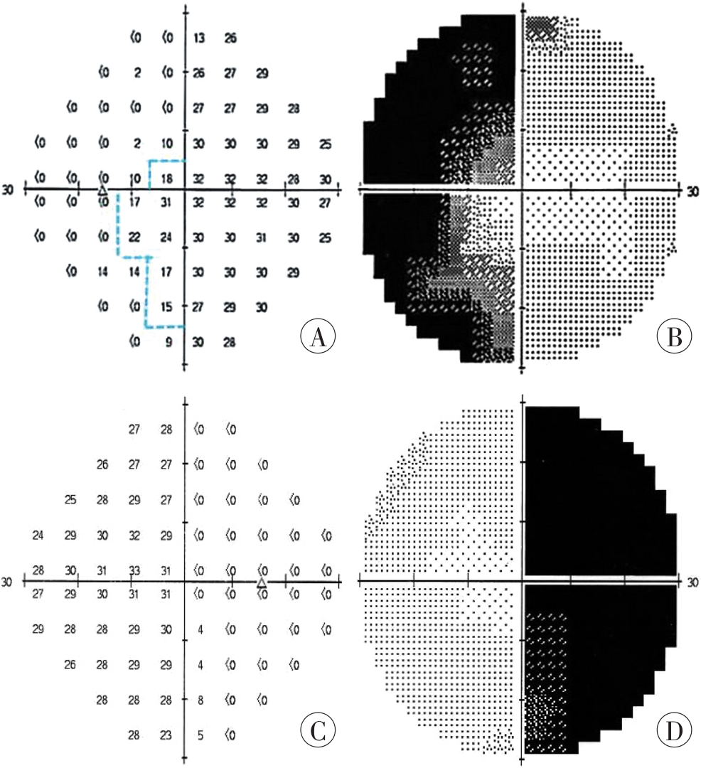
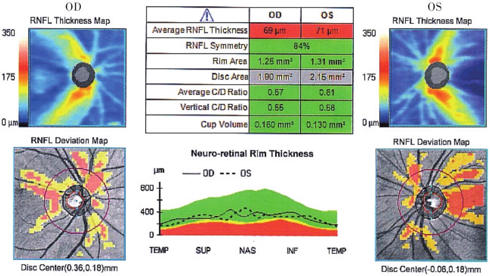
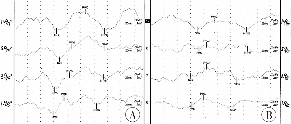
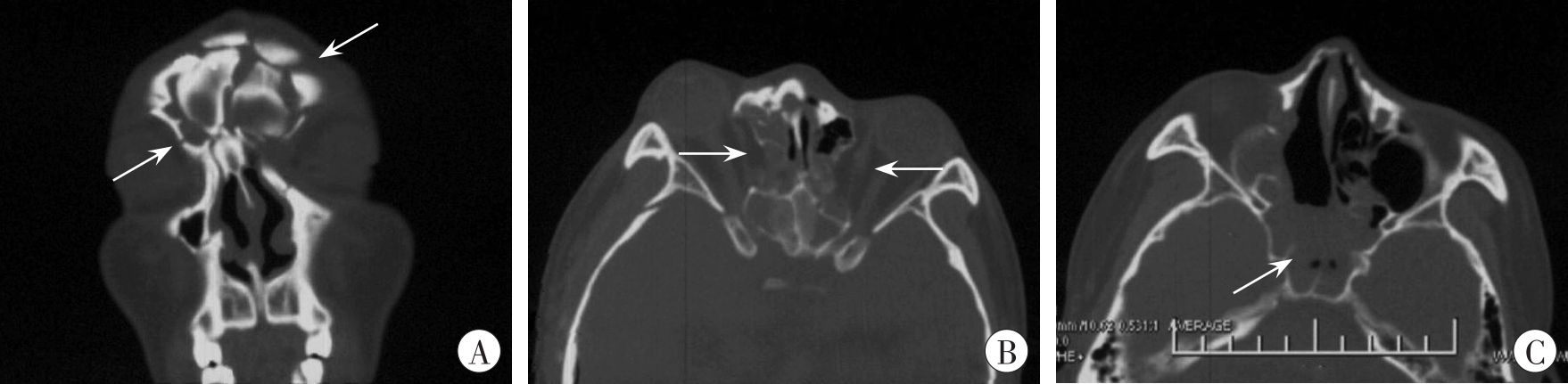
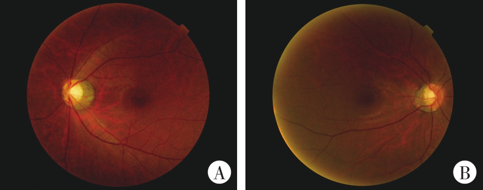
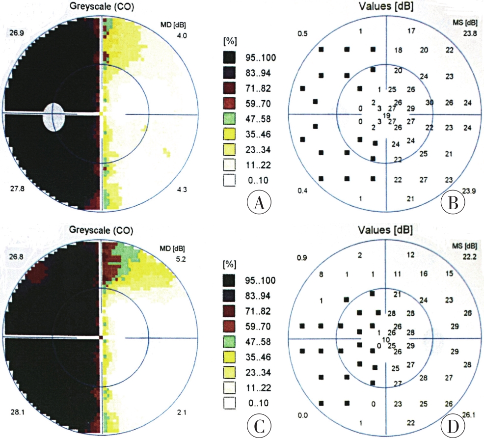
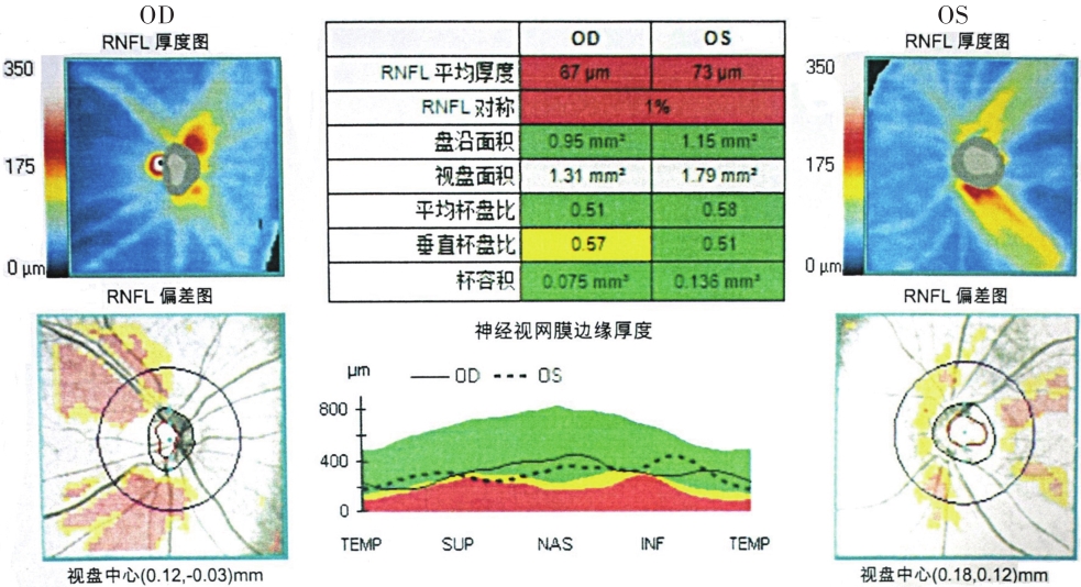
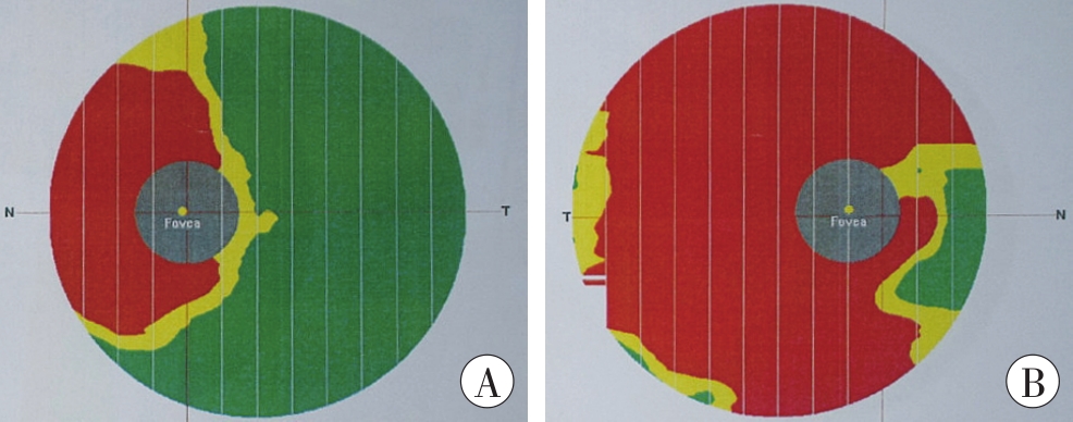
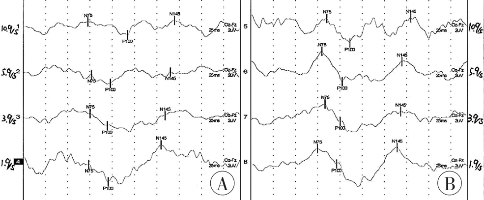
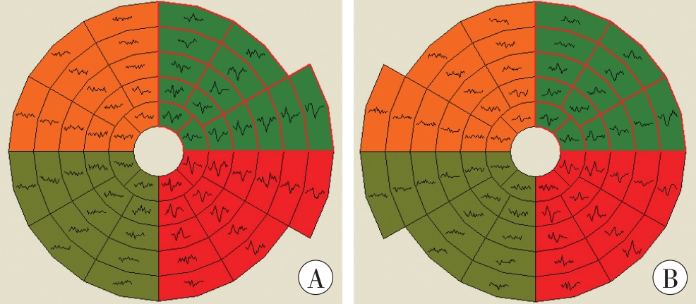
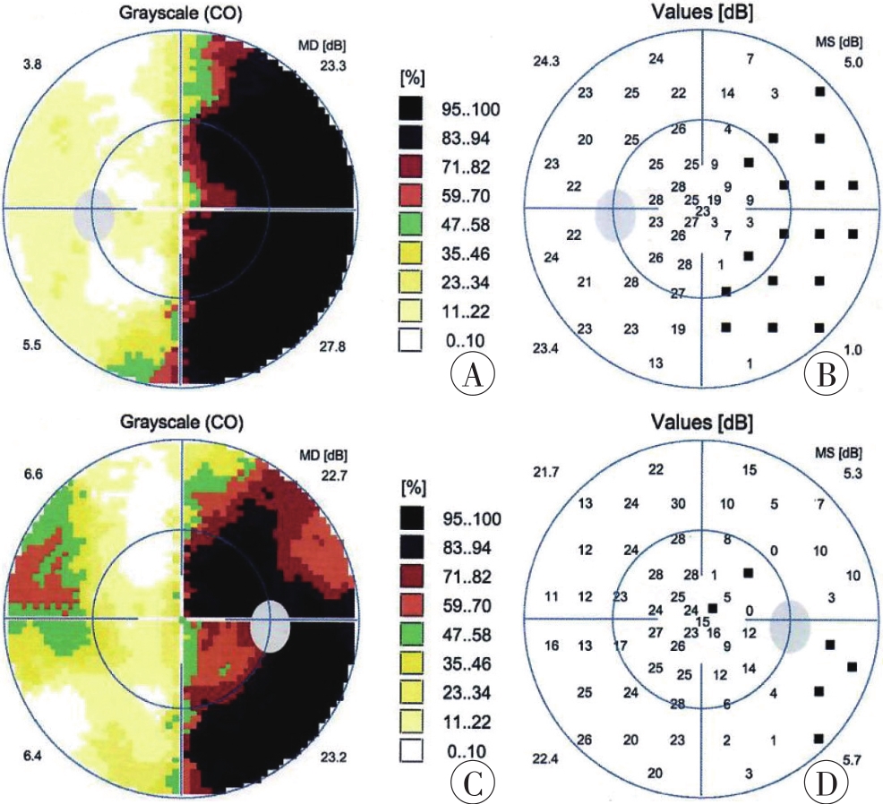
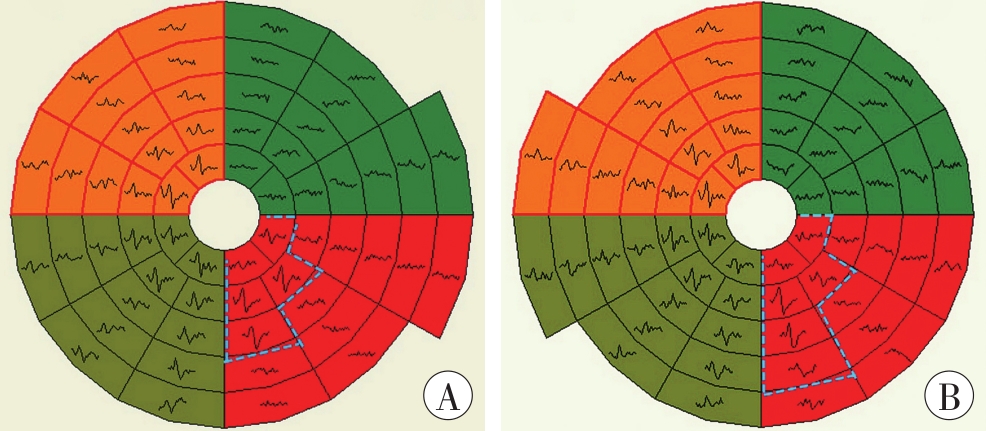
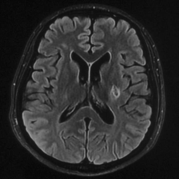
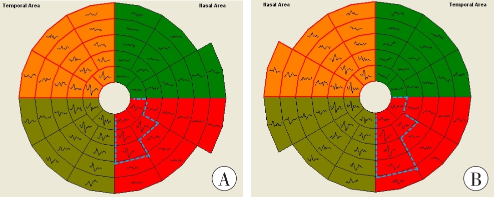
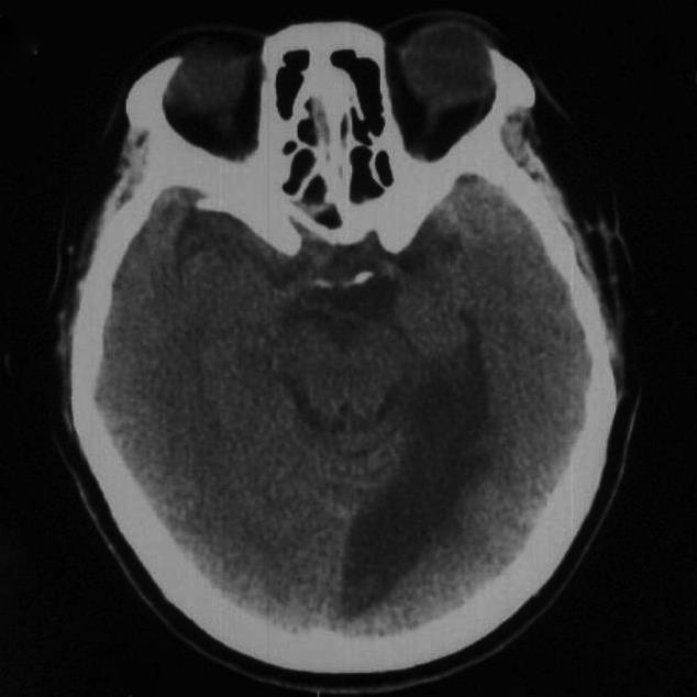
目的 探讨视交叉及其后部视路损伤所致视野缺损的特点及其客观评定方法。 方法 选择视交叉、视束、视放射及视皮质损伤所致视野缺损典型案例,分别进行视野、视觉诱发电位(visual evoked potential,VEP)、多焦视觉诱发电位(multifocal visual evoked potential,mfVEP)检查以及颅脑CT或MRI、视网膜光学相干断层扫描(optical coherence tomography,OCT),并对上述视觉电生理指标及神经影像学指标进行综合分析。 结果 视交叉损伤所致视野缺损的电生理学表现为双颞侧偏盲型mfVEP异常;视束、视放射及视皮质损伤所致视野缺损均表现为病变对侧的同向性偏盲型mfVEP异常。视束损伤出现偏盲眼轻度相对性传入性瞳孔障碍(relative afferent pupil disorder,RAPD)及特征性视神经萎缩表现,视放射、视皮质损伤则无此表现。神经影像学可为视交叉及其后部视路损伤提供形态学证据。 结论 视交叉、视束、视放射及视皮质损伤所致视野缺损具有各自特点,通过mfVEP视野与静态视野检查的联合应用,结合神经影像学检查,可最大程度地评价视路受损的部位和程度,为此类损伤的认定提供有效方案。

关键词: 法医学, 创伤和损伤, 视觉电生理, 视路, 视野, 视交叉, 多焦视觉诱发电位, 视神经损伤

Abstract:

Objective To investigate the characteristics and objective assessment method of visual field defects caused by optic chiasm and its posterior visual pathway injury. Methods Typical cases of visual field defects caused by injuries to the optic chiasm, optic tracts, optic radiations, and visual cortex were selected. Visual field examinations, visual evoked potential (VEP) and multifocal visual evolved potential (mfVEP) measurements, craniocerebral CT/MRI, and retinal optical coherence tomography (OCT) were performed, respectively, and the aforementioned visual electrophysiological and neuroimaging indicators were analyzed comprehensively. Results The electrophysiological manifestations of visual field defects caused by optic chiasm injuries were bitemporal hemianopsia mfVEP abnormalities. The visual field defects caused by optic tract, optic radiation, and visual cortex injuries were all manifested homonymous hemianopsia mfVEP abnormalities contralateral to the lesion. Mild relative afferent pupil disorder (RAPD) and characteristic optic nerve atrophy were observed in hemianopsia patients with optic tract injuries, but not in patients with optic radiation or visual cortex injuries. Neuroimaging could provide morphological evidence of damages to the optic chiasm and its posterior visual pathway. Conclusion Visual field defects caused by optic chiasm, optic tract, optic radiation, and visual cortex injuries have their respective characteristics. The combined application of mfVEP and static visual field measurements, in combination with neuroimaging, can maximize the assessment of the location and degree of visual pathway damage, providing an effective scheme for the identification of such injuries.

Key words: forensic medicine, wounds and injuries, visual electrophysiology, visual pathway, field of view, optic chiasma, multifocal visual evoked potential (mfVEP), optic nerve injury

中图分类号: