Journal of Forensic Medicine ›› 2022, Vol. 38 ›› Issue (1): 46-52.DOI: 10.12116/j.issn.1004-5619.2021.410903
Special Issue: 水中尸体研究专题
• Original Articles • Previous Articles Next Articles
Ji CHEN1( ), Xiao-rong LIU2, Jia-wen YANG1, Ye-qiu CHEN2, Cheng WANG2, Meng-yuan OU2, Jia-yi WU1, You-jia YU1, Kai LI1, Peng CHEN1, Feng CHEN1()
Received:2021-09-01
Online:2022-02-25
Published:2022-02-28
Contact:
Feng CHEN
CLC Number:
Ji CHEN, Xiao-rong LIU, Jia-wen YANG, Ye-qiu CHEN, Cheng WANG, Meng-yuan OU, Jia-yi WU, You-jia YU, Kai LI, Peng CHEN, Feng CHEN. Construction and Application of YOLOv3-Based Diatom Identification Model of Scanning Electron Microscope Images[J]. Journal of Forensic Medicine, 2022, 38(1): 46-52.
Add to citation manager EndNote|Ris|BibTeX
URL: http://www.fyxzz.cn/EN/10.12116/j.issn.1004-5619.2021.410903
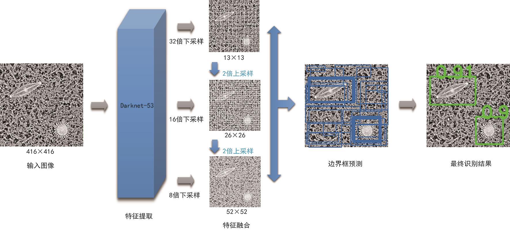
Fig. 1 The schematic diagram of the YOLOv3 model
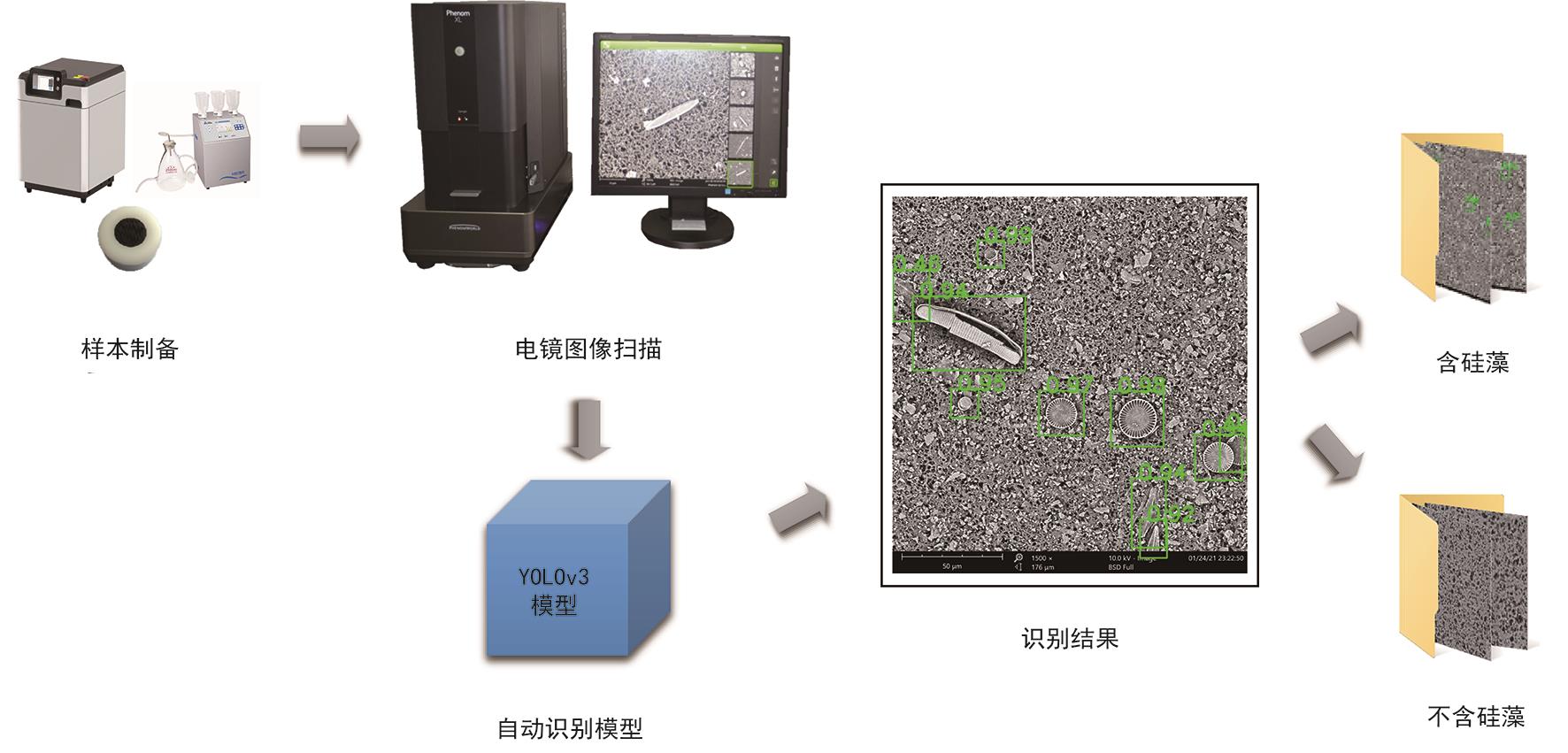
Fig. 2 The flow chart of the diatom identification model of scanning electron microscope images
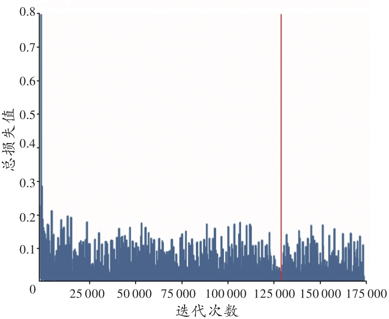
Fig. 3 Convergence curve of model training
| 阈值 | 验证集 | 测试集 | ||||
|---|---|---|---|---|---|---|
| RR/% | PR/% | F1分数 | RR/% | PR/% | F1分数 | |
| 0.05 | 99.2 | 58.9 | 0.739 | 99.5 | 57.6 | 0.729 |
| 0.15 | 98.8 | 79.2 | 0.879 | 98.8 | 76.1 | 0.859 |
| 0.25 | 98.8 | 80.5 | 0.887 | 98.6 | 77.4 | 0.867 |
| 0.35 | 97.9 | 81.4 | 0.889 | 97.2 | 78.1 | 0.866 |
| 0.45 | 77.9 | 95.1 | 0.856 | 77.5 | 92.9 | 0.845 |
| 0.55 | 73.2 | 99.3 | 0.843 | 73.6 | 98.6 | 0.843 |
| 0.65 | 72.4 | 99.6 | 0.838 | 72.9 | 99.5 | 0.841 |
| 0.75 | 71.8 | 99.7 | 0.835 | 72.3 | 99.8 | 0.839 |
| 0.85 | 70.3 | 99.8 | 0.825 | 70.8 | 100.0 | 0.829 |
| 0.95 | 51.4 | 100.0 | 0.679 | 53.3 | 100.0 | 0.695 |
Tab. 1 The precision rate, recall rate and F1 score of the identification model under different thresholds
| 阈值 | 验证集 | 测试集 | ||||
|---|---|---|---|---|---|---|
| RR/% | PR/% | F1分数 | RR/% | PR/% | F1分数 | |
| 0.05 | 99.2 | 58.9 | 0.739 | 99.5 | 57.6 | 0.729 |
| 0.15 | 98.8 | 79.2 | 0.879 | 98.8 | 76.1 | 0.859 |
| 0.25 | 98.8 | 80.5 | 0.887 | 98.6 | 77.4 | 0.867 |
| 0.35 | 97.9 | 81.4 | 0.889 | 97.2 | 78.1 | 0.866 |
| 0.45 | 77.9 | 95.1 | 0.856 | 77.5 | 92.9 | 0.845 |
| 0.55 | 73.2 | 99.3 | 0.843 | 73.6 | 98.6 | 0.843 |
| 0.65 | 72.4 | 99.6 | 0.838 | 72.9 | 99.5 | 0.841 |
| 0.75 | 71.8 | 99.7 | 0.835 | 72.3 | 99.8 | 0.839 |
| 0.85 | 70.3 | 99.8 | 0.825 | 70.8 | 100.0 | 0.829 |
| 0.95 | 51.4 | 100.0 | 0.679 | 53.3 | 100.0 | 0.695 |
| 组织 | 阈值 | 完整硅藻 | 碎片硅藻 | 总体 | ||||||
|---|---|---|---|---|---|---|---|---|---|---|
| RR/% | PR/% | F1分数 | RR/% | PR/% | F1分数 | RR/% | PR/% | F1分数 | ||
| 肺 | 0.4 | 94.0±6.7 | 85.9±27.9 | 0.862±0.225 | 75.8±12.9 | 78.2±29.7 | 0.732±0.219 | 89.6±6.6 | 87.8±25.0 | 0.865±0.181 |
| 0.6 | 81.5±16.1 | 88.4±24.5 | 0.812±0.175 | 39.1±15.0 | 79.6±29.1 | 0.463±0.162 | 69.7±11.8 | 89.8±21.9 | 0.760±0.130 | |
| 0.8 | 71.5±16.8 | 90.0±22.8 | 0.774±0.178 | 24.5±8.3 | 80.2±29.0 | 0.345±0.121 | 58.4±11.8 | 91.0±20.8 | 0.699±0.139 | |
| 肝 | 0.4 | 100.0±0.0 | 1.1±1.2 | 0.022±0.024 | 100.0±0.0 | 1.7±1.2 | 0.034±0.023 | 100.0±0.0 | 2.5±2.0 | 0.049±0.039 |
| 0.6 | 94.4±13.6 | 1.2±1.1 | 0.024±0.021 | 92.7±13.7 | 1.9±1.4 | 0.037±0.027 | 92.3±10.9 | 2.8±2.2 | 0.053±0.039 | |
| 0.8 | 66.7±42.2 | 1.5±1.6 | 0.029±0.030 | 76.0±34.3 | 3.4±2.7 | 0.064±0.050 | 75.6±27.7 | 4.4±3.4 | 0.082±0.061 | |
| 肾 | 0.4 | 100.0±0.0 | 1.7±2.0 | 0.033±0.039 | 100.0±0.0 | 2.4±3.2 | 0.047±0.057 | 100.0±0.0 | 3.6±4.6 | 0.067±0.078 |
| 0.6 | 100.0±0.0 | 2.0±1.9 | 0.039±0.036 | 91.0±17.6 | 2.8±2.9 | 0.053±0.052 | 93.8±11.8 | 4.2±4.3 | 0.077±0.073 | |
| 0.8 | 83.3±40.8 | 3.6±4.3 | 0.066±0.077 | 80.6±22.1 | 4.7±6.1 | 0.085±0.099 | 80.3±26.5 | 6.8±8.5 | 0.118±0.129 | |
Tab. 2 The recall rate, precision rate, and F1 score of the identification model under different thresholds in different tissues(n=8,xˉ±s)
| 组织 | 阈值 | 完整硅藻 | 碎片硅藻 | 总体 | ||||||
|---|---|---|---|---|---|---|---|---|---|---|
| RR/% | PR/% | F1分数 | RR/% | PR/% | F1分数 | RR/% | PR/% | F1分数 | ||
| 肺 | 0.4 | 94.0±6.7 | 85.9±27.9 | 0.862±0.225 | 75.8±12.9 | 78.2±29.7 | 0.732±0.219 | 89.6±6.6 | 87.8±25.0 | 0.865±0.181 |
| 0.6 | 81.5±16.1 | 88.4±24.5 | 0.812±0.175 | 39.1±15.0 | 79.6±29.1 | 0.463±0.162 | 69.7±11.8 | 89.8±21.9 | 0.760±0.130 | |
| 0.8 | 71.5±16.8 | 90.0±22.8 | 0.774±0.178 | 24.5±8.3 | 80.2±29.0 | 0.345±0.121 | 58.4±11.8 | 91.0±20.8 | 0.699±0.139 | |
| 肝 | 0.4 | 100.0±0.0 | 1.1±1.2 | 0.022±0.024 | 100.0±0.0 | 1.7±1.2 | 0.034±0.023 | 100.0±0.0 | 2.5±2.0 | 0.049±0.039 |
| 0.6 | 94.4±13.6 | 1.2±1.1 | 0.024±0.021 | 92.7±13.7 | 1.9±1.4 | 0.037±0.027 | 92.3±10.9 | 2.8±2.2 | 0.053±0.039 | |
| 0.8 | 66.7±42.2 | 1.5±1.6 | 0.029±0.030 | 76.0±34.3 | 3.4±2.7 | 0.064±0.050 | 75.6±27.7 | 4.4±3.4 | 0.082±0.061 | |
| 肾 | 0.4 | 100.0±0.0 | 1.7±2.0 | 0.033±0.039 | 100.0±0.0 | 2.4±3.2 | 0.047±0.057 | 100.0±0.0 | 3.6±4.6 | 0.067±0.078 |
| 0.6 | 100.0±0.0 | 2.0±1.9 | 0.039±0.036 | 91.0±17.6 | 2.8±2.9 | 0.053±0.052 | 93.8±11.8 | 4.2±4.3 | 0.077±0.073 | |
| 0.8 | 83.3±40.8 | 3.6±4.3 | 0.066±0.077 | 80.6±22.1 | 4.7±6.1 | 0.085±0.099 | 80.3±26.5 | 6.8±8.5 | 0.118±0.129 | |
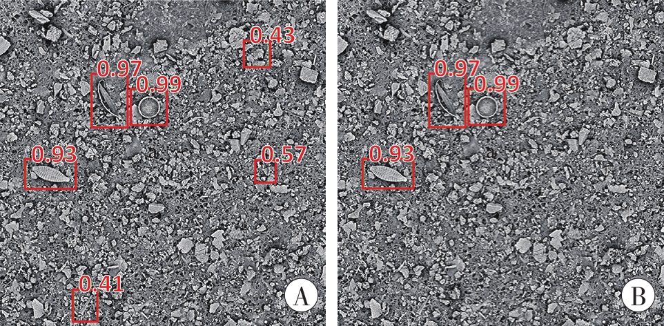
Fig. 4 Diatom identification results under different thresholds
| 1 | 刘超. 溺死法医诊断学[M].广州:中山大学出版社,2018:52-53. |
| LIU C. Diagnosis of drowning[M]. Guangzhou: Sun Yat-sen University Press,2018:52-53. | |
| 2 | PACHAR J V, CAMERON J M. The diagnosis of drowning by quantitative and qualitative diatom analysis[J]. Med Sci Law,1993,33(4):291-299. doi:10.1177/002580249303300404 . |
| 3 | LUDES B, COSTE M, NORTH N, et al. Diatom analysis in victim’s tissues as an indicator of the site of drowning[J]. Int J Legal Med,1999,112(3):163-166. doi:10.1007/s004140050224 . |
| 4 | 李峥,杨佳雯,陈冶秋,等. 硅藻检验推测溺亡位置1例[J].法医学杂志,2020,36(6):878-880. doi:10.12116/j.issn.1004-5619.2020.06.024 . |
| LI Z, YANG J W, CHEN Y Q, et al. Diatom test speculated the location of drowning: A case report[J]. Fayixue Zazhi,2020,36(6):878-880. | |
| 5 | 赵建,袁自闯,张彦吉,等. 两种硅藻检验方法的比较[J].中国法医学杂志,2015,30(1):62-65. doi:10.13618/j.issn.1001-5728.2015.01.018 . |
| ZHAO J, YUAN Z C, ZHANG Y J, et al. The comparative study of two diatom test methods[J]. Zhongguo Fayixue Zazhi,2015,30(1):62-65. | |
| 6 | ZHAO J, LIU C, HU S, et al. Microwave digestion - vacuum filtration - automated scanning electron microscopy as a sensitive method for forensic diatom test[J]. Int J Legal Med,2013,127(2):459-463. doi:10.1007/s00414-012-0756-9 . |
| 7 | LI Y, HUANG Z, DONG X, et al. Forensic age estimation for pelvic X-ray images using deep learning[J]. Eur Radiol,2019,29(5):2322-2329. doi:10.1007/s00330-018-5791-6 . |
| 8 | CHANDRA S S, BRAN LORENZANA M, LIU X, et al. Deep learning in magnetic resonance image reconstruction[J]. J Med Imaging Radiat Oncol,2021,65(5):564-577. doi:10.1111/1754-9485.13276 . |
| 9 | YANG H B, LIU P, HU Y Z, et al. Research on underwater object recognition based on YOLOv3[J]. Microsyst Technol,2021,27(4):1837-1844. doi:10.1007/s00542-019-04694-8 . |
| 10 | ZHOU Y Y, ZHANG J, HUANG J, et al. Digital whole-slide image analysis for automated diatom test in forensic cases of drowning using a convolutional neural network algorithm[J]. Forensic Sci Int,2019,302:109922. doi:10.1016/j.forsciint.2019. 109922 . |
| 11 | 周圆圆,曹永杰,杨越,等. 基于人工智能硅藻自动化识别系统的实际案例应用[J].法医学杂志,2020,36(2):239-242. doi:10.12116/j.issn.1004-5619.2020.02.017 . |
| ZHOU Y Y, CAO Y J, YANG Y, et al. Application of artificial intelligence automatic diatom identification system in practical cases[J]. Fayixue Zazhi,2020,36(2):239-242. | |
| 12 | YU W M, XUE Y, KNOOPS R, et al. Automated diatom searching in the digital scanning electron microscopy images of drowning cases using the deep neural networks[J]. Int J Leg Med,2021,135(2):497-508. doi:10.1007/s00414-020-02392-z . |
| 13 | REDMON J, DIVVALA S, GIRSHICK R, et al. You only look once: Unified, real-time object detection[C]//2016 IEEE Conf Comput Vis Pattern Recognit. Las Vegas: IEEE,2016:779-788. doi:10.1109/CVPR. 2016.91 . |
| 14 | REDMON J, FARHADI A. YOLO9000: Better, faster, stronger[C]//2017 IEEE Conference on Computer Vision and Pattern Recognition. Honolulu: IEEE,2017:6517-6525. doi:10.1109/CVPR.2017.690 . |
| 15 | REDMON J, FARHADI A. YOLOv3: An incremental improvement[J/OL]. (2018-04-08)[2021-08-25]. arXiv:. |
| 16 | 邓杰航,何冬冬,卓家鸿,等. 复杂背景干扰下硅藻图像的深度网络识别与定位[J].南方医科大学学报,2020,40(2):183-189. doi:10.12122/j.issn.1673-4254.2020.02.08 . |
| DENG J H, HE D D, ZHUO J H, et al. Deep learning network-based recognition and localization of diatom images against complex background[J]. Nanfang Yike Daxue Xuebao,2020,40(2):183-189. | |
| 17 | PEDRAZA A, BUENO G, DENIZ O, et al. Automated diatom classification (Part B): A deep learning approach[J]. Appl Sci,2017,7(5):460. doi:10.3390/app7050460 . |
| 18 | KLOSTER M, LANGENKÄMPER D, ZUROWIETZ M, et al. Deep learning-based diatom taxonomy on virtual slides[J]. Sci Rep,2020,10:14416. doi:10.1038/s41598-020-71165-w . |
| 19 | DENG J H, GUO W Q, ZHAO Y W, et al. Identification of diatom taxonomy by a combination of region-based full convolutional network,online hard example mining,and shape priors of diatoms[J]. Int J Leg Med,2021,135(6):2519-2530. doi:10.1007/s00414-021-02664-2 . |
| [1] | Yong ZENG, Dong-hua ZOU, Ying FAN, Qing XU, Lu-yang TAO, Yi-jiu CHEN, Zheng-dong LI. Research Progress and Forensic Application of Human Vascular Finite Element Modeling and Biomechanics [J]. Journal of Forensic Medicine, 2023, 39(5): 471-477. |
| [2] | Yu-xin SUN, Xiao-juan GONG, Xiu-li HAO, Yu-xin TIAN, Yi-ming CHEN, Bao ZHANG, Chun-xia YAN. Screening of Genes Co-Associated with Sudden Infant Death Syndrome and Infectious Sudden Death in Infancy and Bioinformatics Analysis of Their Regulatory Networks [J]. Journal of Forensic Medicine, 2023, 39(5): 433-440. |
| [3] | Yu YANG, Fan-zhang LEI, Yu-you DONG, Jian-long MA, Qi-qiang SHI, Xue-song YE. Retrospective Analysis of Death Cases of Oral Diphenidol Hydrochloride Poisoning [J]. Journal of Forensic Medicine, 2023, 39(4): 393-398. |
| [4] | Jie BAI, Jing SUN, Xiao-guang CHENG, Fan LIU, Hua LIU, Xu WANG. Construction and Application of Rib Fracture Diagnosis Model Based on YOLOv3 Algorithm [J]. Journal of Forensic Medicine, 2023, 39(4): 343-349. |
| [5] | Qing-qing XIANG, Li-fang CHEN, Qin SU, Yu-kun DU, Pei-yan LIANG, Xiao-dong KANG, He SHI, Qu-yi XU, Jian ZHAO, Chao LIU, Xiao-hui CHEN. Research Progress on Microbial Community Succession in the Postmortem Interval Estimation [J]. Journal of Forensic Medicine, 2023, 39(4): 399-405. |
| [6] | Qin SU, Qian-ling CHEN, Wei-bin WU, Qing-qing XIANG, Cheng-liang YANG, Dong-fang QIAO, Zhi-gang LI. Metabonomics Analysis of Brain Stem Tissue in Rats with Primary Brain Stem Injury Caused Death [J]. Journal of Forensic Medicine, 2023, 39(4): 373-381. |
| [7] | Xu-dong ZHANG, Yao-ru JIANG, Xin-rui LIANG, Tian TIAN, Qian-qian JIN, Xiao-hong ZHANG, Jie CAO, Qiu-xiang DU, Jun-hong SUN. Postmortem Interval Estimation Using Protein Chip Technology Combined with Multivariate Analysis Methods [J]. Journal of Forensic Medicine, 2023, 39(2): 115-120. |
| [8] | Yong-gang MA, Yong-jie CAO, Yi-hua ZHAO, Xin-jun ZHOU, Bin HUANG, Gao-chao ZHANG, Ping HUANG, Ya-hui WANG, Kai-jun MA, Feng CHEN, Dong-chuan ZHANG, Ji ZHANG. Sex Estimation of Medial Aspect of the Ischiopubic Ramus in Adults Based on Deep Learning [J]. Journal of Forensic Medicine, 2023, 39(2): 129-136. |
| [9] | Wu LONG, Peng-fei QU, Lin MA, Rui WANG, Yan-mei XI, Yu-hua LI, Sheng-jie NIE, Ting DUAN, Jin-liang DU, Xue TANG, Jing-feng ZHAO, Pu-ping LEI, Yue-bing WANG. Cytotoxicity of 4 Wild Mushrooms in a Case of Yunnan Sudden Unexplained Death [J]. Journal of Forensic Medicine, 2023, 39(2): 121-128. |
| [10] | Jun-wei GAO, Yang LU, Yan-jun LI, Dong-hua ZOU, Guang-long HE, Yan-bin WANG. Survey on the Construction Status of Forensic Virtual Autopsy Laboratory and the Applicability of Laboratory Accreditation [J]. Journal of Forensic Medicine, 2023, 39(2): 186-192. |
| [11] | Fang-Fang LIU, Hui WU, Wei WANG, Ying XIE. Changes of 1,5-AG in Vitreous Humor of Rabbit Cadavers with Hyperglycemic Metabolism [J]. Journal of Forensic Medicine, 2023, 39(1): 13-17. |
| [12] | Zhi-ling TIAN, Ruo-lin WANG, Jian-hua ZHANG, Ping HUANG, Zhi-qiang QIN, Zheng-dong LI, He-wen DONG, Dong-hua ZOU, Mao-wen WANG, Zhuo LI, Lei WAN, Xiao-tian YU, Ning-guo LIU. Comparison of CT Values between Thrombus and Postmortem Clot Based on Cadaveric Pulmonary Angiography [J]. Journal of Forensic Medicine, 2023, 39(1): 7-12. |
| [13] | Hong-xia HAO, Ya-hui WANG, Zhi-lu ZHOU, Tai-ang LIU, Jin CHEN, Yu-heng HE, Lei WAN, Wen-tao XIA. Research Progress of Age Estimation in the Living by Knee Joint MRI [J]. Journal of Forensic Medicine, 2023, 39(1): 66-71. |
| [14] | Jia-yi WU, You-jia YU, Kai LI, Xin YIN, Han-ting FAN, Rong LI, Zhi-wei ZHANG, Wei TANG, Hui-jie HUANG, Feng CHEN. Report on Cardiac Gross Pathologic Measurements of Sudden Cardiac Death in Adults [J]. Journal of Forensic Medicine, 2023, 39(1): 1-6. |
| [15] | Hui WU, Fang-fang LIU, Jun-da WU, Ying XIE. Research Progress on Estimation of Postmortem Interval Based on Ocular Tissues Structure [J]. Journal of Forensic Medicine, 2023, 39(1): 50-56. |
| Viewed | ||||||
|
Full text |
|
|||||
|
Abstract |
|
|||||