Journal of Forensic Medicine ›› 2023, Vol. 39 ›› Issue (6): 535-541.DOI: 10.12116/j.issn.1004-5619.2023.430317
• Original Articles •
Tian TIAN1,2( ), Xin-biao LIAO3, Fu ZHANG3, Kai-fei DENG1, Ji ZHANG1, Ping HUANG1, Yi-jiu CHEN1, Jian-hua ZHANG1()
Received:2023-03-26
Online:2024-01-17
Published:2023-12-25
Contact:
Jian-hua ZHANG
CLC Number:
Tian TIAN, Xin-biao LIAO, Fu ZHANG, Kai-fei DENG, Ji ZHANG, Ping HUANG, Yi-jiu CHEN, Jian-hua ZHANG. Forensic Pathological Diagnosis of Acute and Old Myocardial Infarction Using Fourier Transform Infrared Spectroscopy[J]. Journal of Forensic Medicine, 2023, 39(6): 535-541.
Add to citation manager EndNote|Ris|BibTeX
URL: http://www.fyxzz.cn/EN/10.12116/j.issn.1004-5619.2023.430317

Fig. 1 Pathological changes of the control myocardium and early ischemic myocardium
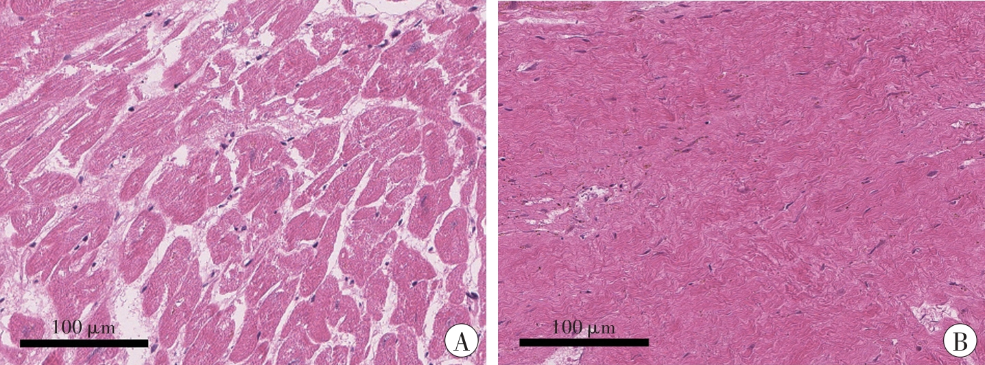
Fig. 2 Pathological changes of the necrotic myocardium and myocardial fibrous tissue (HE staining)

Fig. 3 The mean normalized spectra and the mean second derivative spectra of different types of myocardium

Fig. 4 PCA scores plot, PC2 loading plot and PLS-DA scores plot of the second derivative spectra ofdifferent types of myocardium

Fig. 5 PCA scores plot, PC2 loading plot and PLS-DA scores plot of the second derivative spectra ofcontrol myocardium, early ischemic myocardium and necrotic myocardium

Fig. 6 PCA scores plots and PLS-DA scores plots for two-by-two subgroups of the second derivative spectra ofcontrol myocardium, early ischemic myocardium and necrotic myocardium
| 1 | MARIJON E, GARCIA R, NARAYANAN K, et al. Fighting against sudden cardiac death: Need for a paradigm shift -- Adding near-term prevention and pre-emptive action to long-term prevention[J]. Eur Heart J,2022,43(15):1457-1464. doi:10.1093/eurheartj/ehab903 . |
| 2 | LOU J, CHEN H, HUANG S, et al. Update on risk factors and biomarkers of sudden unexplained cardiac death[J]. J Forensic Leg Med,2022,87:102332. doi:10.1016/j.jflm.2022.102332 . |
| 3 | ALPERT J S, THYGESEN K, ANTMAN E, et al. Myocardial infarction redefined -- A consensus document of The Joint European Society of Cardiology/American College of Cardiology Committee for the redefinition of myocardial infarction[J]. J Am Coll Cardiol,2000,36(3):959-969. doi:10.1016/s0735-1097 (00)00804-4 . |
| 4 | TANG W H W, FRANCIS G S, MORROW D A, et al. National academy of clinical biochemistry laboratory medicine practice guidelines: Clinical utilization of cardiac biomarker testing in heart failure[J]. Circulation,2007,116(5):e99-109. doi:10 . |
| 1161/CIRCULATIONAHA.107.185267. | |
| 5 | SABATASSO S, MORETTI M, MANGIN P, et al. Early markers of myocardial ischemia: From the experimental model to forensic pathology cases of sudden cardiac death[J]. Int J Legal Med,2018,132(1):197-203. doi:10.1007/s00414-017-1605-7 . |
| 6 | ELLIS D I, GOODACRE R. Metabolic fingerprinting in disease diagnosis: Biomedical applications of infrared and Raman spectroscopy[J]. Analyst,2006,131(8):875-885. doi:10.1039/b602376m . |
| 7 | CHEHELTANI R, ROSANO J M, WANG B, et al. Fourier transform infrared spectroscopic imaging of cardiac tissue to detect collagen deposition after myocardial infarction[J]. J Biomed Opt,2012,17(5):056014. doi:10.1117/1.JBO.17.5.056014 . |
| 8 | LIN H, WANG Z, LUO Y, et al. Post-mortem evaluation of the pathological degree of myocardial infarction by Fourier transform infrared microspectroscopy[J]. Spectrochim Acta A Mol Biomol Spectrosc,2022,268:120630. doi:10.1016/j.saa.2021.120630 . |
| 9 | TIAN T, ZHANG J, XIONG L, et al. Evaluating subtle pathological changes in early myocardial ische-mia using spectral histopathology[J]. Anal Chem,2022,94(49):17112-17120. doi:10.1021/acs.analchem.2c03368 . |
| 10 | BYLER D M, SUSI H. Examination of the secondary structure of proteins by deconvolved FTIR spectra[J]. Biopolymers,1986,25(3):469-487. doi:10.1002/bip.360250307 . |
| 11 | MONDELLO C, CARDIA L, VENTURA-SPAGNO-LO E. Immunohistochemical detection of early myocardial infarction: A systematic review[J]. Int J Legal Med,2017,131(2):411-421. doi:10.1007/s00414-016-1494-1 . |
| 12 | BELBACHIR K, NOREEN R, GOUSPILLOU G, et al. Collagen types analysis and differentiation by FTIR spectroscopy[J]. Anal Bioanal Chem,2009,395(3):829-837. doi:10.1007/s00216-009-3019-y . |
| 13 | COLLINSON P O, GAZE D C. Biomarkers of cardiovascular damage[J]. Med Princ Pract,2007,16(4):247-261. doi:10.1159/000102146 . |
| 14 | SABATASSO S, MANGIN P, FRACASSO T, et al. Early markers for myocardial ischemia and sudden cardiac death[J]. Int J Legal Med,2016,130(5):1265-1280. doi:10.1007/s00414-016-1401-9 . |
| 15 | MONDELLO C, VENTURA SPAGNOLO E, CAR-DIA L, et al. Membrane attack complex in myocardial ischemia/reperfusion injury: A systematic review for post mortem applications[J]. Diagnostics (Basel),2020,10(11):898. doi:10.3390/diagnostics10110898 . |
| 16 | 丛璐,蒲传强. 膜攻击复合物C5b-9的研究现状[J].中华医学杂志,2012,92(1):67-70. doi:10.3760/cma.j.issn.0376-2491.2012.01.019 . |
| CONG L, PU C Q. Research status of membrane attack complex C5b-9[J]. Zhonghua Yixue Zazhi,2012,92(1):67-70. | |
| 17 | BROMBERG P S, GOUGH K M, DIXON I M C. Collagen remodeling in the extracellular matrix of the cardiomyopathic Syrian hamster heart as assessed by FTIR attenuated total reflectance spectroscopy[J]. Can J Chem,1999,77(11):1843-1855. doi:10.1139/v99-178 . |
| 18 | ZHENG N, YANG T, LIANG M, et al. Characterization of protein in old myocardial infarction by FTIR micro-spectroscopy[J]. J Huazhong Univ Sci Technolog Med Sci,2010,30(4):546-550. doi:10.1007/s11596-010-0466-9 . |
| 19 | ALDOUS S J. Cardiac biomarkers in acute myocardial infarction[J]. Int J Cardiol,2013,164(3):282-294. doi:10.1016/j.ijcard.2012.01.081 . |
| 20 | GOFF K L, ELLIS T H, WILSON K E. Synchrotron FTIR spectromicroscopy as a tool for studying populations and individual living cells of green algae[J]. Analyst,2021,145(24):7993-8001. doi:10.1039/d0an01386b . |
| 21 | 黄平,黎世莹,李正东,等. 傅里叶显微红外光谱成像技术研究进展及其法医学应用[J].法医学杂志,2011,27(6):447-450. doi:10.3969/j.issn.1004-5619.2011.06.013 . |
| HUANG P, LI S Y, LI Z D, et al. Research advancement of FTIR-MSP mapping and application value in forensic science[J]. Fayixue Zazhi,2011,27(6):447-450. | |
| 22 | CAMPOBASSO C P, DELL’ERBA A S, ADDANTE A, et al. Sudden cardiac death and myocardial ischemia indicators: A comparative study of four immunohistochemical markers[J]. Am J Forensic Med Pathol,2008,29(2):154-161. doi:10.1097/paf.0b013e318177eab7 . |
| 23 | CALABRÒ E, CONDELLO S, CURRÒ M, et al. 50 Hz electromagnetic field produced changes in FTIR spectroscopy associated with mitochondrial transmembrane potential reduction in neuronal-like SH-SY5Y cells[J]. Oxid Med Cell Longev,2013,2013:414393. doi:10.1155/2013/414393 . |
| 24 | NEVRAUMONT A, DELTOMBE M, FAVRESSE J, et al. Interferences with cardiac biomarker assays: Understanding the clinical impact[J]. Eur Heart J,2022,43(24):2286-2288. doi:10.1093/eurheartj/ehab924 . |
| [1] | Yong ZENG, Dong-hua ZOU, Ying FAN, Qing XU, Lu-yang TAO, Yi-jiu CHEN, Zheng-dong LI. Research Progress and Forensic Application of Human Vascular Finite Element Modeling and Biomechanics [J]. Journal of Forensic Medicine, 2023, 39(5): 471-477. |
| [2] | Yu-xin SUN, Xiao-juan GONG, Xiu-li HAO, Yu-xin TIAN, Yi-ming CHEN, Bao ZHANG, Chun-xia YAN. Screening of Genes Co-Associated with Sudden Infant Death Syndrome and Infectious Sudden Death in Infancy and Bioinformatics Analysis of Their Regulatory Networks [J]. Journal of Forensic Medicine, 2023, 39(5): 433-440. |
| [3] | Yu YANG, Fan-zhang LEI, Yu-you DONG, Jian-long MA, Qi-qiang SHI, Xue-song YE. Retrospective Analysis of Death Cases of Oral Diphenidol Hydrochloride Poisoning [J]. Journal of Forensic Medicine, 2023, 39(4): 393-398. |
| [4] | Qing-qing XIANG, Li-fang CHEN, Qin SU, Yu-kun DU, Pei-yan LIANG, Xiao-dong KANG, He SHI, Qu-yi XU, Jian ZHAO, Chao LIU, Xiao-hui CHEN. Research Progress on Microbial Community Succession in the Postmortem Interval Estimation [J]. Journal of Forensic Medicine, 2023, 39(4): 399-405. |
| [5] | Qin SU, Qian-ling CHEN, Wei-bin WU, Qing-qing XIANG, Cheng-liang YANG, Dong-fang QIAO, Zhi-gang LI. Metabonomics Analysis of Brain Stem Tissue in Rats with Primary Brain Stem Injury Caused Death [J]. Journal of Forensic Medicine, 2023, 39(4): 373-381. |
| [6] | Xu-dong ZHANG, Yao-ru JIANG, Xin-rui LIANG, Tian TIAN, Qian-qian JIN, Xiao-hong ZHANG, Jie CAO, Qiu-xiang DU, Jun-hong SUN. Postmortem Interval Estimation Using Protein Chip Technology Combined with Multivariate Analysis Methods [J]. Journal of Forensic Medicine, 2023, 39(2): 115-120. |
| [7] | Wu LONG, Peng-fei QU, Lin MA, Rui WANG, Yan-mei XI, Yu-hua LI, Sheng-jie NIE, Ting DUAN, Jin-liang DU, Xue TANG, Jing-feng ZHAO, Pu-ping LEI, Yue-bing WANG. Cytotoxicity of 4 Wild Mushrooms in a Case of Yunnan Sudden Unexplained Death [J]. Journal of Forensic Medicine, 2023, 39(2): 121-128. |
| [8] | Jun-wei GAO, Yang LU, Yan-jun LI, Dong-hua ZOU, Guang-long HE, Yan-bin WANG. Survey on the Construction Status of Forensic Virtual Autopsy Laboratory and the Applicability of Laboratory Accreditation [J]. Journal of Forensic Medicine, 2023, 39(2): 186-192. |
| [9] | Fang-Fang LIU, Hui WU, Wei WANG, Ying XIE. Changes of 1,5-AG in Vitreous Humor of Rabbit Cadavers with Hyperglycemic Metabolism [J]. Journal of Forensic Medicine, 2023, 39(1): 13-17. |
| [10] | Zhi-ling TIAN, Ruo-lin WANG, Jian-hua ZHANG, Ping HUANG, Zhi-qiang QIN, Zheng-dong LI, He-wen DONG, Dong-hua ZOU, Mao-wen WANG, Zhuo LI, Lei WAN, Xiao-tian YU, Ning-guo LIU. Comparison of CT Values between Thrombus and Postmortem Clot Based on Cadaveric Pulmonary Angiography [J]. Journal of Forensic Medicine, 2023, 39(1): 7-12. |
| [11] | Jia-yi WU, You-jia YU, Kai LI, Xin YIN, Han-ting FAN, Rong LI, Zhi-wei ZHANG, Wei TANG, Hui-jie HUANG, Feng CHEN. Report on Cardiac Gross Pathologic Measurements of Sudden Cardiac Death in Adults [J]. Journal of Forensic Medicine, 2023, 39(1): 1-6. |
| [12] | Hui WU, Fang-fang LIU, Jun-da WU, Ying XIE. Research Progress on Estimation of Postmortem Interval Based on Ocular Tissues Structure [J]. Journal of Forensic Medicine, 2023, 39(1): 50-56. |
| [13] | Tian-pu WU, Jian-long MA, Xin-biao LIAO, Dong-chuan ZHANG, Kai-jun MA, Yan-geng YU, Long CHEN. Research Progress on Molecular Changes in Pulmonary Hypoxia and Cause of Death Identification in Mechanical Asphyxia [J]. Journal of Forensic Medicine, 2023, 39(1): 57-65. |
| [14] | Yu-qing JIA, Tian-qi WANG, Rui ZHAO, Bao-li ZHU, Zhi-peng CAO. Effects of Postmortem Hemolysis and Ultrafiltration on Creatinine Detection Results [J]. Journal of Forensic Medicine, 2022, 38(6): 697-701. |
| [15] | Lan YANG, Xin WANG, Yong NIU. Research Progress of DNA-Based Technologies for Postmortem Interval Estimation [J]. Journal of Forensic Medicine, 2022, 38(6): 747-753. |
| Viewed | ||||||
|
Full text |
|
|||||
|
Abstract |
|
|||||