法医学杂志 ›› 2025, Vol. 41 ›› Issue (5): 494-501.DOI: 10.12116/j.issn.1004-5619.2024.440115

• 论著 • 上一篇    

铁死亡在小鼠百草枯急性肺损伤中的作用

秦豪杰1,2(), 莫晨1, 李洪伟1, 刘之江3, 郑哲1,2   

  1. 1.河南科技大学基础医学与法医学院,河南 洛阳 471023
    2.河南科技大学司法鉴定中心,河南 洛阳 471023
    3.宁波市公安局鄞州分局,浙江 宁波 315100
  • 收稿日期:2024-01-22 发布日期:2026-01-27 出版日期:2025-10-25
  • 作者简介:秦豪杰(1978—),男,博士,副教授,硕士研究生导师,主要从事法医学教学、科研和鉴定;E-mail:herochin@haust.edu.cn
  • 基金资助:
    河南省科技攻关计划项目(232102310352)

Role of Ferroptosis in Paraquat-Induced Acute Lung Injury in Mice

Hao-jie QIN1,2(), Chen MO1, Hong-wei LI1, Zhi-jiang LIU3, Zhe ZHENG1,2   

  1. 1.College of Basic Medicine and Forensic Medicine, Henan University of Science and Technology, Luoyang 471023, Henan Province, China
    2.Forensic Medicine Identification Center of Henan University of Science and Technology, Luoyang 471023, Henan Province, China
    3.Yinzhou Branch of Ningbo Public Security Bureau, Ningbo 315100, Zhejiang Province, China
  • Received:2024-01-22 Online:2026-01-27 Published:2025-10-25

摘要:

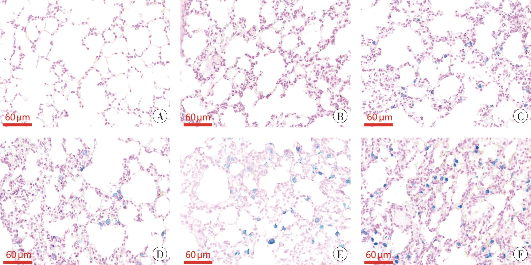
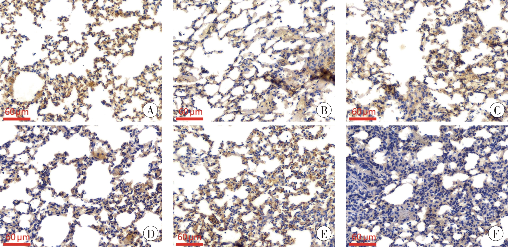
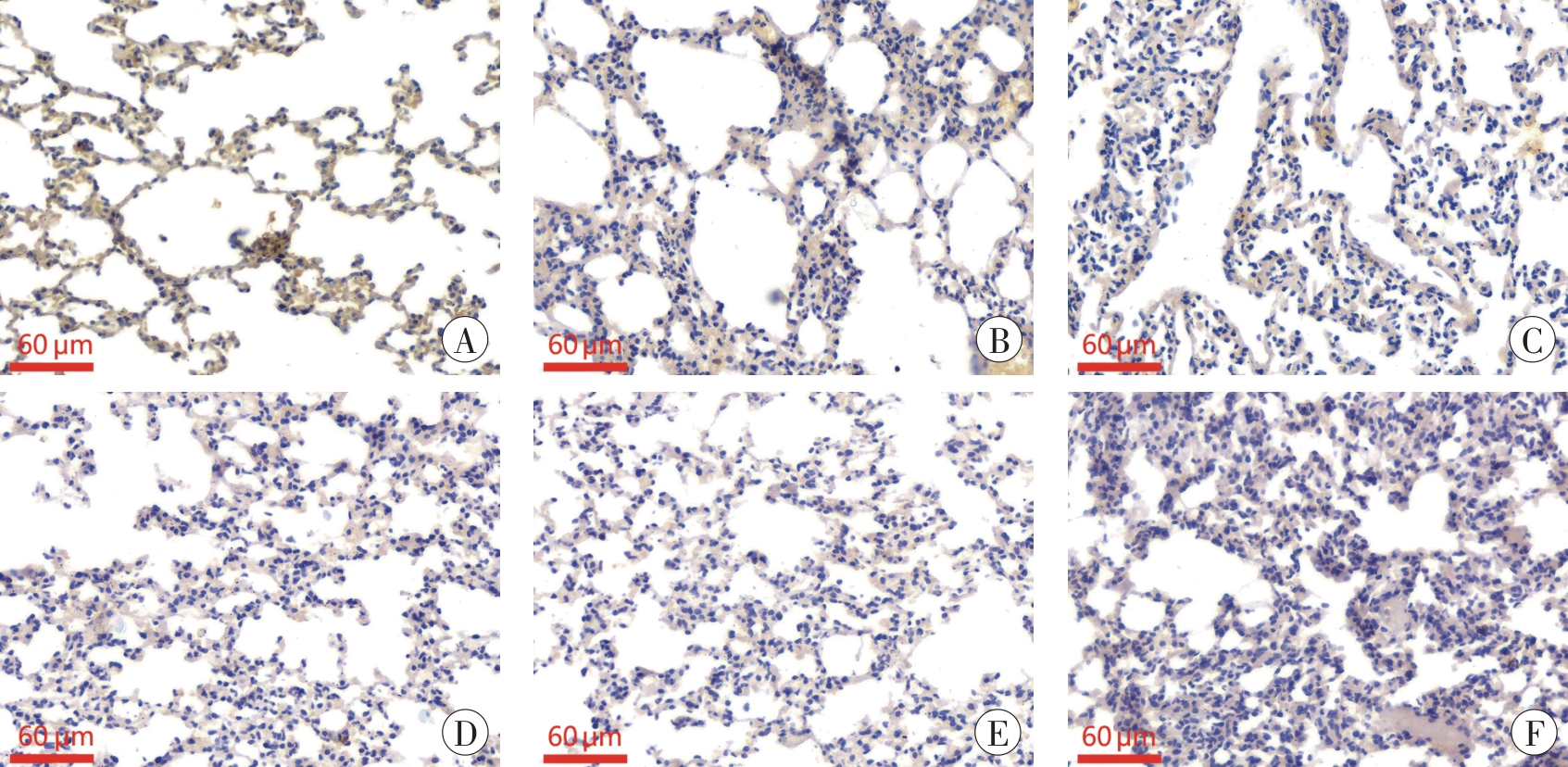
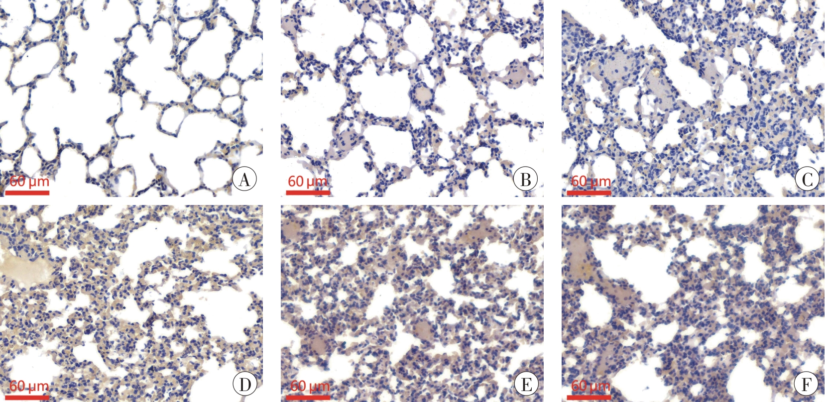
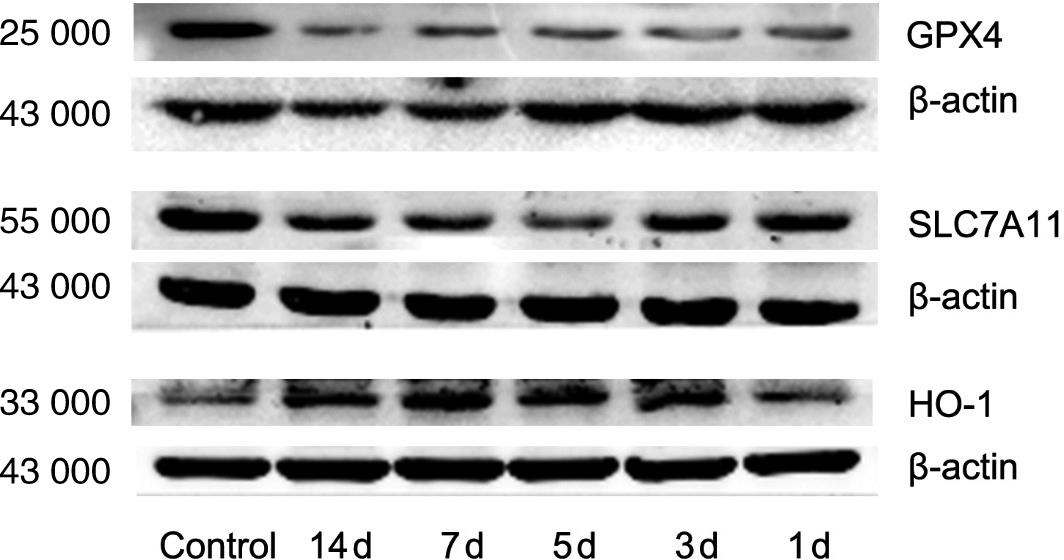
目的 探究铁死亡在小鼠百草枯急性肺损伤中可能的作用机制及法医学意义。 方法 选取6~8周龄清洁级雄性昆明小鼠60只,随机分为对照组、染毒1 d组、染毒3 d组、染毒5 d组、染毒7 d组和染毒14 d组,每组10只。染毒组小鼠一次性腹腔注射20 mg/kg百草枯纯品溶液,对照组一次性腹腔注射20 mg/kg 0.9%生理盐水。观察小鼠染毒后一般状态,采用HE染色观察肺组织病理学改变,普鲁士蓝染色观测肺组织铁含量,试剂盒检测还原型谷胱甘肽(glutathione, GSH)、丙二醛(malondialdehyde, MDA)等氧化应激指标,免疫组织化学法和蛋白质印迹法检测肺组织铁死亡相关蛋白谷胱甘肽过氧化物酶4(glutathione peroxidase 4, GPX4)、溶质载体家族7成员11(solute carrier family 7 member 11, SLC7A11)及血红素氧合酶-1(heme oxygenase-1, HO-1)的表达情况。 结果 随染毒时间增加,染毒组小鼠肺组织进行性纤维化加重,铁沉积增多;GSH含量逐渐下降,MDA含量于染毒7 d组和14 d组增加;肺组织GPX4蛋白表达下降;SLC7A11蛋白除染毒1 d组外,随染毒时间增加表达下降;HO-1蛋白除染毒1 d组外,随染毒时间增加表达增高,染毒7 d组表达最高,差异均有统计学意义(P<0.05)。 结论 铁死亡参与百草枯中毒急性肺损伤,对铁死亡机制及相关指标时序性变化的研究可望为百草枯中毒的法医学鉴定提供思路。

关键词: 法医病理学, 法医毒理学, 百草枯, 急性肺损伤, 细胞铁死亡, 小鼠

Abstract:

Objective To explore the potential mechanisms by which ferroptosis contributes to paraquat-induced acute lung injury in mice and its forensic significance. Methods Sixty healthy male clean-grade Kunming mice aged 6-8 weeks were randomly divided into a control group and paraquat exposure groups (1 d, 3 d, 5 d, 7 d and 14 d), with 10 mice per group. The exposure groups were given a single intraperitoneal injection of 20 mg/kg paraquat solution, while the control group was given an equal volume of 0.9% saline. General conditions after exposure were observed. Hematoxylin-eosin (HE) staining was used to examine pathological changes in lung tissues, and Prussian blue staining was used to detect iron content. Oxidative stress indicators, such as reduced glutathione (GSH) and malondialdehyde (MDA), were measured by relevant testing kits. Immunohistochemistry and Western blot were employed to detect the expression levels of ferroptosis-related proteins including glutathione peroxidase 4 (GPX4), solute carrier family 7 member 11(SLC7A11), and heme oxygenase-1 (HO-1). Results With the increase of exposure time, mice in the exposure groups showed progressively aggravated fibrosis and increased iron deposition in lung tissues. GSH levels gradually decreased, while MDA levels increased significantly in the 7 d and 14 d groups. The expression of GPX4 in lung tissue decreased. The expression of SLC7A11 decreased over time, except for the 1 d group. The expression of HO-1 increased with the increase of exposure time, except for the 1 d group, and the highest expression was observed in the 7 d group. The differences between the exposure groups and the control group were statistically significant (P<0.05). Conclusion Ferroptosis is involved in acute lung injury induced by paraquat poisoning, and research on the mechanism of ferroptosis and the temporal changes of its biomarkers may provide valuable insights for the forensic identification of paraquat poisoning.

Key words: forensic pathology, forensic toxicology, paraquat, acute lung injury, ferroptosis, mice

中图分类号: