法医学杂志 ›› 2025, Vol. 41 ›› Issue (2): 144-151.DOI: 10.12116/j.issn.1004-5619.2024.440503

• 论著中毒的法医学鉴定专题 • 上一篇    下一篇

钙调控蛋白与核因子κB在急性MDMA染毒心肌细胞钙失衡中的作用机制

王荣帅1,2(), 黄锶哲1,3, 王云云1, 邓燕飞1,4, 丁自娇1,5, 张杰1, 刘勇1, 任亮1(), 刘良1()   

  1. 1.华中科技大学同济医学院法医学系,湖北 武汉 430030
    2.皖南医学院法医学院,安徽 芜湖 241000
    3.南方医科大学第七附属医院,广东 佛山 528244
    4.江西省公安厅,江西 南昌 330038
    5.安徽医科大学基础医学院法医学系,安徽 合肥 230032
  • 发布日期:2025-08-11 出版日期:2025-04-25
  • 通讯作者: 任亮,刘良
  • 作者简介:王荣帅(1986—),男,讲师,主要从事法医病理学和法医毒理学研究;E-mail:wangrs87@126.com
  • 基金资助:
    国家自然科学基金资助项目(81373243);皖南医学院中青年科研基金资助项目(WYRCQD2022004);法庭科学湖北省重点实验室开放课题资助项目(KFKT2023009)

The Mechanism of Calcium Handling Proteins and NF-κB in Calcium Dyshomeostasis of Cardiomyocytes Caused by Acute MDMA Exposure

Rong-shuai WANG1,2(), Si-zhe HUANG1,3, Yun-yun WANG1, Yan-fei DENG1,4, Zi-jiao DING1,5, Jie ZHANG1, Yong LIU1, Liang REN1(), Liang LIU1()   

  1. 1.Department of Forensic Medicine, Tongji Medical College, Huazhong University of Science and Technology, Wuhan 430030, China
    2.School of Forensic Medicine, Wannan Medical College, Wuhu 241000, Anhui Province, China
    3.The Seventh Affiliated Hospital of Southern Medical University, Foshan 528244, Guangdong Province, China
    4.Jiangxi Provincial Public Security Department, Nanchang 330038, China
    5.Department of Forensic Medicine, School of Basic Medical Sciences, Anhui Medical University, Hefei 230032, China
  • Online:2025-08-11 Published:2025-04-25
  • Contact: Liang REN, Liang LIU

摘要:

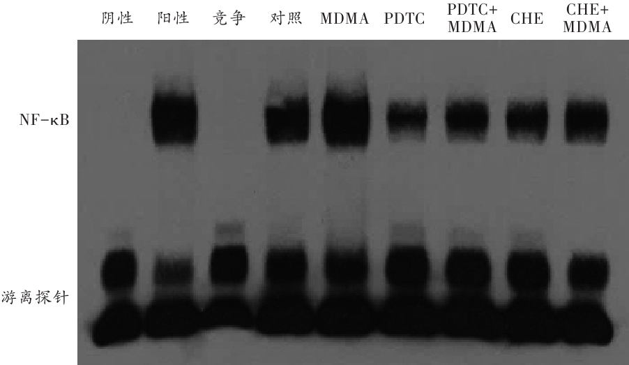
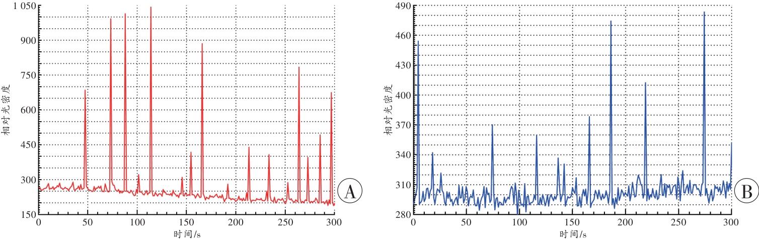
目的 通过检测不同浓度3,4-亚甲二氧基甲基苯丙胺(N-methyl-3,4-methylenedioxyamphetamine,MDMA)急性染毒后心肌细胞内钙振荡模式变化特征和差异、钙调控蛋白变化以及核因子κB(nuclear factor κB,NF-κB)是否参与及其对钙调控蛋白的影响,探讨MDMA致心肌毒性的作用机制。 方法 采用原代大鼠心肌细胞建立急性MDMA染毒模型,并设立对照组。MDMA染毒组分为10、100和1 000 μmol/L 3个浓度组,染毒1 h后观察心肌细胞形态变化,测定细胞毒性及钙信号的变化特征,并检测钙调控蛋白RyR2、SERCA2a、PLN、NCX1及Cav1.2的变化。使用电泳迁移率变动分析(electrophoretic mobility shift assay,EMSA)法和Western印迹法检测MDMA染毒及NF-κB抑制剂吡咯烷二硫代氨基甲酸铵(pyrrolidine dithiocarbamate ammonium,PDTC)、蛋白质激酶C(protein kinase C,PKC)抑制剂白屈菜红碱(chelerythrine,CHE)干预后NF-κB的活性变化和核蛋白p-p65(Ser311)、PKCζ的表达变化,并观察PDTC干预后MDMA染毒对心肌细胞内钙信号的影响及钙调控蛋白RyR2、SERCA2a、PLN、NCX1和Cav1.2的变化。 结果 急性MDMA染毒后心肌细胞形态未见明显改变;细胞质内钙离子振荡波形呈不规则改变,振荡波幅增大,波动剧烈,频率不规则,相对光密度值波动幅度增大;心肌细胞内RyR2、SERCA2a及NCX1表达增加,Cav1.2、PLN表达下降。急性MDMA染毒可增加NF-κB活性,PDTC及CHE干预后可抑制NF-κB活性。在MDMA染毒组中,PKCζ及核蛋白p-p65(Ser311)表达均有所增加,并可被CHE抑制。PDTC干预阻断NF-κB后,染毒心肌细胞内钙振荡波幅较MDMA染毒组降低,RyR2、SERCA2a、NCX1表达下降,PLN无明显变化,而Cav1.2表达增加。 结论 MDMA可以导致心肌细胞内钙离子浓度升高,这一过程参与心肌毒性作用,其机制与心肌细胞内钙调控蛋白的变化有关,主要与RyR2表达增加相关;MDMA可通过PKCζ-NF-κB途径上调细胞内NF-κB活性,并作用于心肌细胞钙调控蛋白,加剧急性MDMA染毒时的细胞内钙离子超载。

关键词: 法医病理学, 法医毒理学, 3,4-亚甲二氧基甲基苯丙胺(MDMA), 细胞毒性, 钙稳态, 钙调控蛋白, 核因子κB, 蛋白质激酶C

Abstract:

Objective To explore the mechanism of myocardial toxicity caused by N-methyl-3,4-methyle-nedioxyamphetamine (MDMA), the changes of intracellular calcium oscillation mode and calcium handling proteins during acute exposure to different concentrations of MDMA were detected, and the involvement of nuclear factor κB (NF-κB) and its effect on calcium handling proteins were investigated. Methods Primary rat cardiomyocytes were cultured to establish MDMA acute exposure model, and a control group was set up. The MDMA poisoning model was divided into three concentration groups of 10, 100 and 1 000 μmol/L. After 1 h of exposure, the morphological changes of cardiomyocytes were observed, the cytotoxicity and changes in calcium signals were measured, and the changes in calcium handling proteins RyR2, SERCA2a, PLN, NCX1 and Cav1.2 were detected. The changes of NF-κB activity and the expression of nucleoprotein p-p65 (Ser311) and PKCζ after MDMA exposure, and the intervention of NF-κB inhibitors pyrrolidine dithiocarbamate ammonium (PDTC) and protein kinase C (PKC) inhibitor chelerythrine (CHE) were detected by electrophoretic mobility shift assay (EMSA) and Western blotting. The effects of PDTC intervention on calcium signals, and the expressions of RyR2, SERCA2a, PLN, NCX1 and Cav1.2 after acute MDMA exposure were also observed. Results No obvious changes were observed in the morphology of cardiomyocytes after acute exposure to MDMA, whereas the oscillation waveform of intracytoplasmic calcium ion showed irregular changes with increased oscillation amplitude, intense fluctuations, irregular frequency, and increased fluctuation range of relative optical density values. The expression of RyR2, SERCA2a and NCX1 increased, while the expression of Cav1.2 and PLN decreased. Acute MDMA exposure could increase NF-κB activity, while PDTC and CHE intervention could inhibit NF-κB activity. In MDMA exposed group, the expression of PKCζ and nucleoprotein p-p65 (Ser311) both increased and could be inhibited by CHE. After the intervention of PDTC to block NF-κB, the amplitude of calcium oscillation was lower than that of the MDMA exposed group, and the expression of RyR2, SERCA2a and NCX1 decreased. There was no significant change in PLN, while the expression of Cav1.2 increased. Conclusion MDMA can lead to an increase of calcium ion concentration in cardiomyocytes. Calcium ions are involved in myocardial toxicity of MDMA. The mechanism is related to changes in calcium handling proteins, mainly associated with the increased expression of RyR2. MDMA can up-regulate the intracellular activity of NF-κB through the PKCζ-NF-κB pathway and affect calcium handling proteins, which aggravate intracellular calcium overload during acute MDMA exposure.

Key words: forensic pathology, forensic toxicology, N-methyl-3,4-methylenedioxyamphetamine (MDMA), cytotoxicity, calcium homeostasis, calcium handling protein, nuclear factor κB (NF-κB), protein kinase C (PKC)

中图分类号: