法医学杂志 ›› 2025, Vol. 41 ›› Issue (2): 152-159.DOI: 10.12116/j.issn.1004-5619.2024.440411

• 论著中毒的法医学鉴定专题 • 上一篇    下一篇

铁死亡在α-鹅膏毒肽诱导肝细胞损伤中的作用

王浩伟(), 张晓星, 杨根梦, 王尚文(), 曾晓锋()   

  1. 昆明医科大学法医学院,云南 昆明 650500
  • 收稿日期:2024-04-19 发布日期:2025-08-11 出版日期:2025-04-25
  • 通讯作者: 王尚文,曾晓锋
  • 作者简介:王浩伟(1998—),男,博士研究生,主要从事毒蘑菇中毒机制研究;E-mail:1300386555@qq.com
  • 基金资助:
    云南省科技厅应用基础研究青年项目(202301AU070216);云南省科技厅-昆明医科大学联合专项(202301AY070001-263);上海市法医学重点实验室暨司法部司法鉴定重点实验室开放课题资助项目(KF202301);昆明医科大学博士研究生教育创新基金资助项目(2025B021)

The Role of Ferroptosis in Hepatocyte Injury Induced by α-Amanitin

Hao-wei WANG(), Xiao-xing ZHANG, Gen-meng YANG, Shang-wen WANG(), Xiao-feng ZENG()   

  1. College of Forensic Medicine, Kunming Medical University, Kunming 650500, China
  • Received:2024-04-19 Online:2025-08-11 Published:2025-04-25
  • Contact: Shang-wen WANG, Xiao-feng ZENG

摘要:

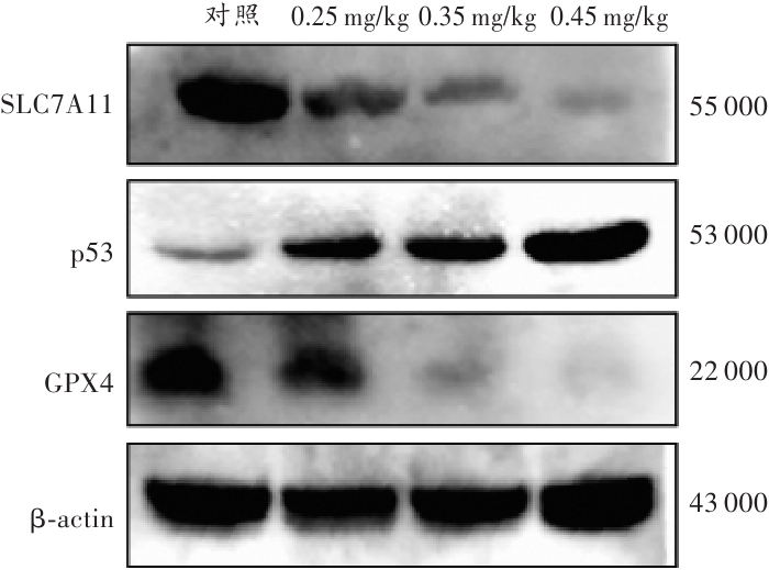
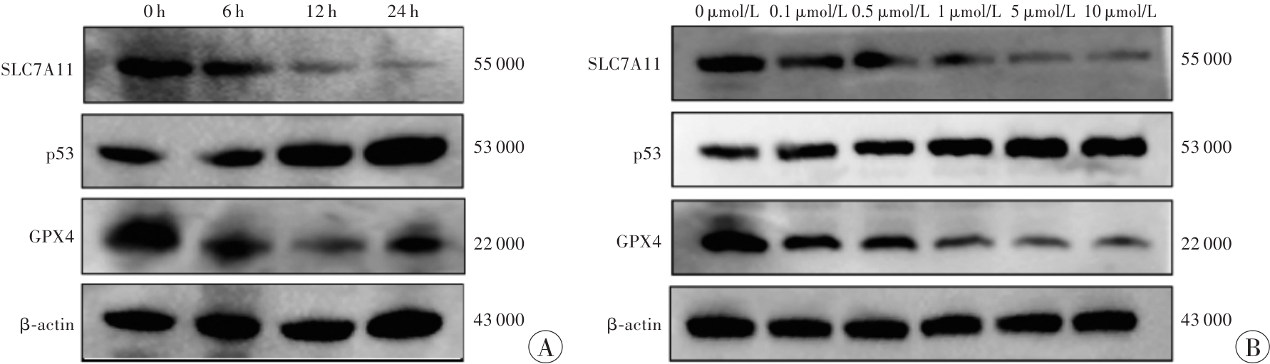
目的 通过检测α-鹅膏毒肽暴露后小鼠肝组织铁沉积,小鼠肝细胞和L-02细胞氧化应激指标及铁死亡相关蛋白的表达,探究铁死亡是否参与α-鹅膏毒肽诱导的肝细胞损伤。 方法 建立α-鹅膏毒肽C57BL/6J小鼠中毒模型和L-02细胞染毒模型,采用Lillie二价铁染色、普鲁士蓝染色检测铁沉积情况,试剂盒检测超氧化物歧化酶(superoxide dismutase,SOD)、过氧化氢酶(catalase,CAT)、丙二醛(malondialdehyde,MDA)、谷胱甘肽(glutathione,GSH)水平,蛋白质印迹法分析p53、溶质载体家族7成员11(solute carrier family 7 member 11,SLC7A11)、谷胱甘肽过氧化物酶4(glutathione peroxidase 4,GPX4)的表达。 结果 相较于对照组,α-鹅膏毒肽暴露后,小鼠肝组织中二价铁(Fe²⁺)和三价铁(Fe³⁺)阳性细胞率均升高(P<0.05),中(0.35 mg/kg)、高(0.45 mg/kg)剂量组肝组织及1 μmol/L α-鹅膏毒肽处理的L-02细胞中GSH水平降低、MDA水平升高,SOD和CAT活性下降(P<0.05)。此外,α-鹅膏毒肽呈浓度和时间依赖性上调p53表达,并抑制SLC7A11和GPX4的表达。 结论 铁死亡在α-鹅膏毒肽诱导的肝细胞损伤机制中发挥作用,铁死亡相关指标的异常可为α-鹅膏毒肽中毒的法医学鉴定提供参考。

关键词: 法医病理学, 法医毒理学, α-鹅膏毒肽, 铁死亡, 氧化应激, 肝损伤, L-02细胞, 小鼠

Abstract:

Objective To explore whether ferroptosis is involved in α-amanitin-induced hepatocyte injury by detecting iron deposition in mice liver tissues, oxidative stress indicators in hepatocytes and L-02 cells, and expressions of ferroptosis-related proteins after α-amanitin exposure. Methods The poisoning models of α-amanitin C57BL/6J mice and L-02 cell were established. The Lillie ferrous iron staining and Prussian blue staining were used to detect iron deposition; the kits were applied to detect the levels of superoxide dismutase (SOD), catalase (CAT), malondialdehyde (MDA), and glutathione (GSH). Western blotting was performed to analyze expressions of p53, solute carrier family 7 member 11 (SLC7A11), and glutathione peroxidase 4 (GPX4). Results Compared with the control group, after α-amanitin exposure, positive cell rates of Fe2+ and Fe3+ in mice liver tissues increased significantly. In the liver tissues of medium (0.35 mg/kg) and high (0.45 mg/kg) dose groups and L-02 cells treated with 1 μmol/L α-amanitin, the level of GSH decreased, the level of MDA increased, and the activities of SOD and CAT decreased significantly. In addition, α-amanitin upregulated the expression of p53 in a concentration- and time-dependent manner and inhibited the expressions of SLC7A11 and GPX4. Conclusion Ferroptosis plays an important role in α-amanitin-induced hepatocyte injury. Abnormalities of ferroptosis-related indicators can provide references for the forensic identification of α-amanitin poisoning.

Key words: forensic pathology, forensic toxicology, α-amanitin, ferroptosis, oxidative stress, hepatic injury, L-02 cell, mice

中图分类号: