Journal of Forensic Medicine ›› 2025, Vol. 41 ›› Issue (5): 494-501.DOI: 10.12116/j.issn.1004-5619.2024.440115
• Original Article • Previous Articles
Hao-jie QIN1,2( ), Chen MO1, Hong-wei LI1, Zhi-jiang LIU3, Zhe ZHENG1,2
Received:2024-01-22
Online:2026-01-27
Published:2025-10-25
CLC Number:
Hao-jie QIN, Chen MO, Hong-wei LI, Zhi-jiang LIU, Zhe ZHENG. Role of Ferroptosis in Paraquat-Induced Acute Lung Injury in Mice[J]. Journal of Forensic Medicine, 2025, 41(5): 494-501.
Add to citation manager EndNote|Ris|BibTeX
URL: http://www.fyxzz.cn/EN/10.12116/j.issn.1004-5619.2024.440115

Fig. 1 Pathological changes of mice lung tissues at different time points after paraquat poisoning
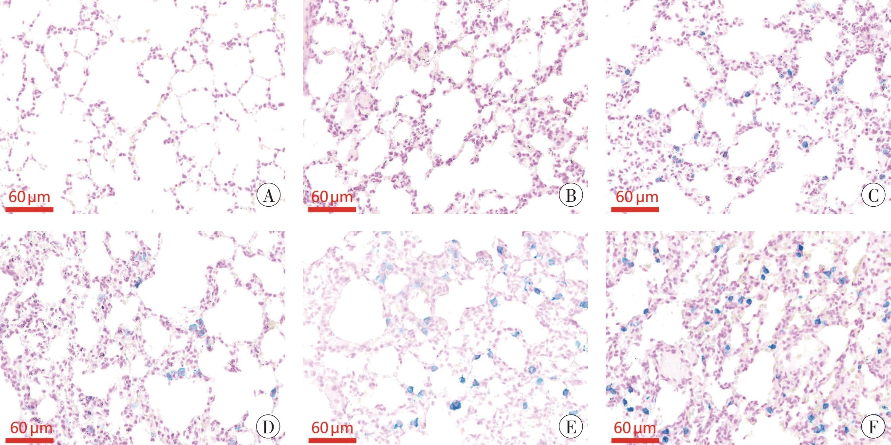
Fig. 2 Iron staining results of mice lung tissues at different time points after paraquat poisoning
| 组别 | GSH/(μg·g-1) | MDA/(nmoL·g-1) |
|---|---|---|
| 对照 | 712.6±5.0 | 45.9±5.0 |
| 染毒1 d | 650.8±6.0 1) | 50.4±4.2 |
| 染毒3 d | 615.9±4.8 1)2) | 51.1±3.3 |
| 染毒5 d | 560.5±13.3 1)2) | 49.7±3.5 |
| 染毒7 d | 521.1±2.7 1)2) | 61.2±9.1 1) |
| 染毒14 d | 460.6±5.3 1)2) | 76.3±11.9 1) |
Tab. 1 The GSH and MDA contents in mice lung tissues
| 组别 | GSH/(μg·g-1) | MDA/(nmoL·g-1) |
|---|---|---|
| 对照 | 712.6±5.0 | 45.9±5.0 |
| 染毒1 d | 650.8±6.0 1) | 50.4±4.2 |
| 染毒3 d | 615.9±4.8 1)2) | 51.1±3.3 |
| 染毒5 d | 560.5±13.3 1)2) | 49.7±3.5 |
| 染毒7 d | 521.1±2.7 1)2) | 61.2±9.1 1) |
| 染毒14 d | 460.6±5.3 1)2) | 76.3±11.9 1) |
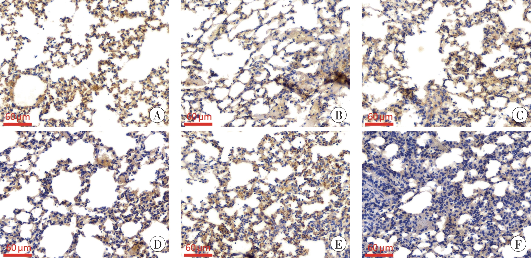
Fig. 3 Immunohistochemical staining results of GPX4 in mice lung tissues at different time points after paraquat poisoning
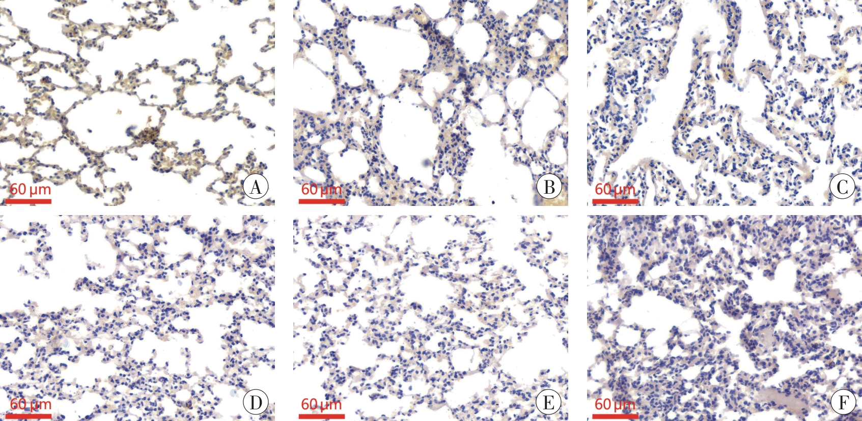
Fig. 4 Immunohistochemical staining results of SLC7A11 in mice lung tissues at different time points after paraquat poisoning
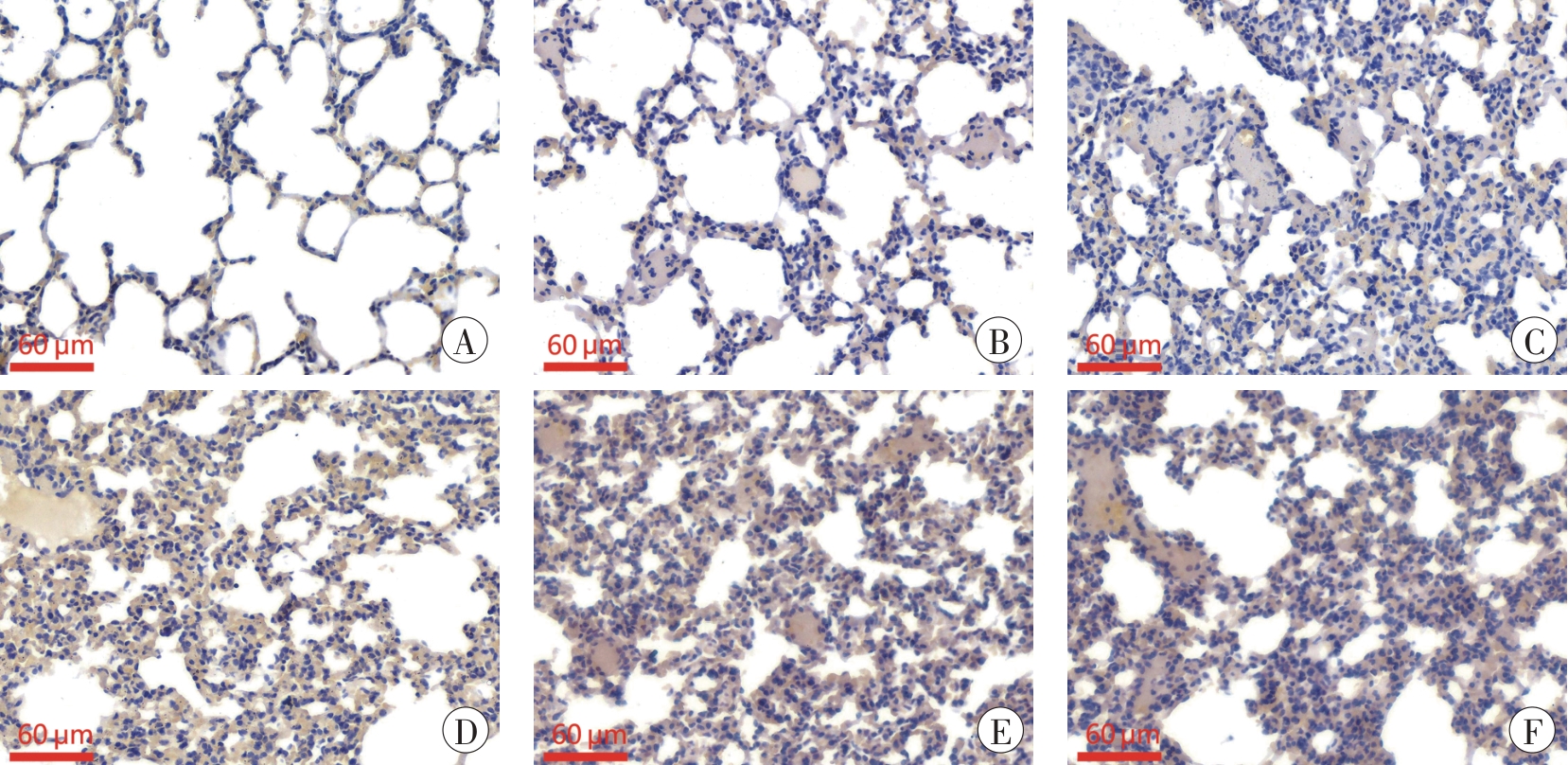
Fig. 5 Immunohistochemical staining results of HO-1 in mice lung tissues at different time points after paraquat poisoning
| 组别 | GPX4 | SLC7A11 | HO-1 |
|---|---|---|---|
| 对照 | 1 | 1 | 1 |
| 染毒1 d | 0.58±0.09 1) | 0.84±0.06 | 1.31±0.06 |
| 染毒3 d | 0.46±0.07 1)2) | 0.66±0.06 1) | 1.40±0.11 1) |
| 染毒5 d | 0.34±0.08 1)2) | 0.64±0.14 1) | 1.55±0.06 1)2) |
| 染毒7 d | 0.35±0.11 1)2) | 0.62±0.07 1)2) | 1.79±0.10 1)2) |
| 染毒14 d | 0.32±0.07 1)2) | 0.58±0.05 1)2) | 1.52±0.06 1)2) |
Tab. 2 Relative expression levels of GPX4, SLC7A11 and HO-1 proteins in mice lung tissues atdifferent time points after paraquat poisoning
| 组别 | GPX4 | SLC7A11 | HO-1 |
|---|---|---|---|
| 对照 | 1 | 1 | 1 |
| 染毒1 d | 0.58±0.09 1) | 0.84±0.06 | 1.31±0.06 |
| 染毒3 d | 0.46±0.07 1)2) | 0.66±0.06 1) | 1.40±0.11 1) |
| 染毒5 d | 0.34±0.08 1)2) | 0.64±0.14 1) | 1.55±0.06 1)2) |
| 染毒7 d | 0.35±0.11 1)2) | 0.62±0.07 1)2) | 1.79±0.10 1)2) |
| 染毒14 d | 0.32±0.07 1)2) | 0.58±0.05 1)2) | 1.52±0.06 1)2) |
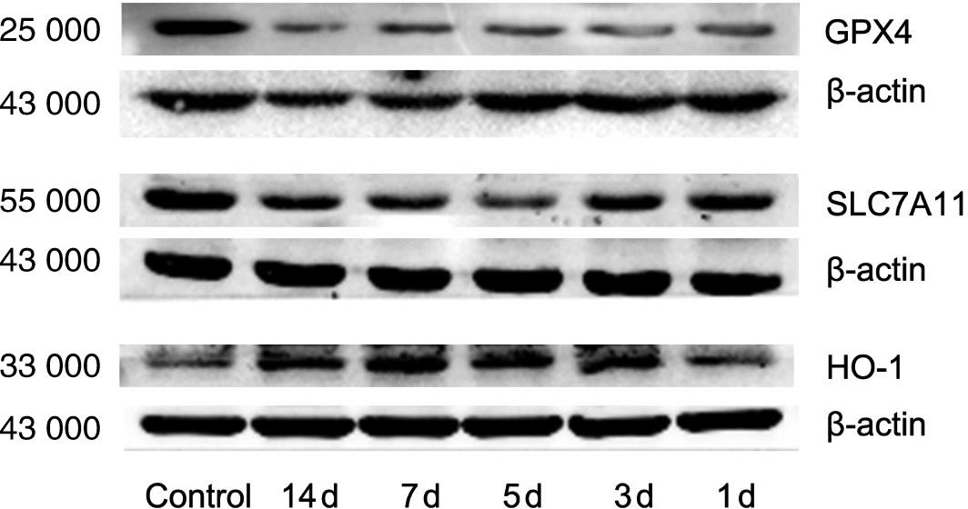
Fig. 6 Changes of GPX4, SLC7A11 and HO-1 protein expression in mice lung tissue at different time points after paraquat poisoning
| [1] | LIU Z, HUANG F, ZHAO S, et al. Homicidal paraquat poisoning: Poisoned while drinking[J]. J Forensic Sci,2022,67(3):1312-1319. doi:10.1111/1556-4029.14968 . |
| [2] | SHI M, ZENG M, JIAN T, et al. A mass event of paraquat poisoning via inhalation[J]. Front Public Health,2023,11:1309708. doi:10.3389/fpubh. 2023.1309708 . |
| [3] | ELENGA N, MERLIN C, LE GUERN R, et al. Clinical features and prognosis of paraquat poisoning in French Guiana: A review of 62 cases[J]. Medicine (Baltimore),2018,97(15):e9621. doi:10.1097/MD.0000000000009621 . |
| [4] | SHADNIA S, EBADOLLAHI-NATANZI A, AHMADZADEH S, et al. Delayed death following paraquat poisoning: Three case reports and a literature review[J]. Toxicol Res (Camb),2018,7(5):745-753. doi:10.1039/c8tx 00120k . |
| [5] | 吉子炎. 百草枯肌肉注射后迟发性死亡1例[J].法医学杂志,2017,33(5):552-553. doi:10.3969/j.issn.1004-5619.2017.05.025 . |
| JI Z Y. Delayed death after intramuscular injection of paraquat: A case report[J]. Fayixue Zazhi,2017,33(5):552-553. | |
| [6] | 刘勇,王方,赵波,等. 隐匿性百草枯中毒1例[J].法医学杂志,2016,32(2):154-155. doi:10.3969/j.issn.1004-5619.2016.02.025 . |
| LIU Y, WANG F, ZHAO B, et al. Occult paraquat poisoning: A case report[J]. Fayixue Zazhi,2016,32(2):154-155. | |
| [7] | DIXON S J, LEMBERG K M, LAMPRECHT M R, et al. Ferroptosis: An iron-dependent form of nonapoptotic cell death[J]. Cell,2012,149(5):1060-1072. doi:10.1016/j.cell.2012.03.042 . |
| [8] | DIXON S J, PATEL D N, WELSCH M, et al. Pharmacological inhibition of cystine-glutamate exchange induces endoplasmic reticulum stress and ferroptosis[J]. Elife,2014,3:e02523. doi:10.7554/eLife.02523 . |
| [9] | KANER R J, LADETTO J V, SINGH R, et al. Lung overexpression of the vascular endothelial growth factor gene induces pulmonary edema[J]. Am J Respir Cell Mol Biol,2000,22(6):657-664. doi:10.1165/ajrcmb.22.6.3779 . |
| [10] | HENNINGSSON R, ALM P, LUNDQUIST I. Evaluation of islet heme oxygenase-CO and nitric oxide synthase-NO pathways during acute endotoxemia[J]. Am J Physiol Cell Physiol,2001,280(5):C1242-C1254. doi:10.1152/ajpcell.2001.280.5.C1242 . |
| [11] | 刘建辉,李欣,孙志平,等. 百草枯中毒所致大鼠急性肺损伤时肺内HO-1/CO的变化[J].中国煤炭工业医学杂志,2006,9(5):511-512. doi:10.3969/j.issn.1007-9564.2006.05.083 . |
| LIU J H, LI X, SUN Z P, et al. Changes of HO-1/CO in the lungs of levels in rats with acute lung injury induced by paraquat poisoning[J]. Zhongguo Meitan Gongye Yixue Zazhi,2006,9(5):511-512. | |
| [12] | CHANG L C, CHIANG S K, CHEN S E, et al. Heme oxygenase-1 mediates BAY 11-7085 induced ferroptosis[J]. Cancer Lett,2018,416:124-137. doi:10.1016/j.canlet.2017.12.025 . |
| [13] | TSIKAS D. Assessment of lipid peroxidation by measuring malondialdehyde (MDA) and relatives in biological samples: Analytical and biological challenges[J]. Anal Biochem,2017,524:13-30. doi:10.1016/j.ab.2016.10.021 . |
| [14] | 王萍,李铁刚. 百草枯中毒致小鼠肺纤维化模型的建立[J].实用药物与临床,2020,23(10):886-889. doi:10.14053/j.cnki.ppcr.202010003 . |
| WANG P, LI T G. Establishment of a mouse lung fibrosis model induced by paraquat poisoning[J]. Shiyong Yaowu Yu Linchuang,2020,23(10):886-889. | |
| [15] | LIU Z, WANG X, LI L, et al. Hydrogen sulfide protects against paraquat-induced acute liver injury in rats by regulating oxidative stress, mitochondrial function, and inflammation[J]. Oxid Med Cell Longev,2020,2020:6325378. doi:10.1155/2020/6325378 . |
| [16] | SUN B, CHEN Y G. Advances in the mechanism of paraquat-induced pulmonary injury[J]. Eur Rev Med Pharmacol Sci,2016,20(8):1597-1602. |
| [17] | FENG H, STOCKWELL B R. Unsolved mysteries: How does lipid peroxidation cause ferroptosis?[J]. PLoS Biol, 2018,16(5):e2006203. doi:10.1371/journal.pbio.2006203 . |
| [18] | YANG W S, STOCKWELL B R. Ferroptosis: Death by lipid peroxidation[J]. Trends Cell Biol,2016,26(3):165-176. doi:10.1016/j.tcb.2015.10.014 . |
| [19] | KOPPULA P, ZHANG Y, ZHUANG L, et al. Amino acid transporter SLC7A11/xCT at the crossroads of regulating redox homeostasis and nutrient dependency of cancer[J]. Cancer Commun (Lond),2018,38(1):12. doi:10.1186/s40880-018-0288-x . |
| [20] | SUN L, DONG H, ZHANG W, et al. Lipid peroxidation, GSH depletion, and SLC7A11 inhibition are common causes of EMT and ferroptosis in A549 cells, but different in specific mechanisms[J]. DNA Cell Biol, 2021,40(2):172-183. doi:10.1089/dna.2020.5730 . |
| [21] | CHIANG S K, CHEN S E, CHANG L C. A dual role of heme oxygenase-1 in cancer cells[J]. Int J Mol Sci,2018,20(1):39. doi:10.3390/ijms 20010039 . |
| [22] | 龙伟,徐丽华,成燕,等. 铁死亡参与小鼠脓毒血症相关急性肺损伤的形成[J].西南国防医药,2020,30(8):725-728. doi:10.3969/j.issn.1004-0188.2020.08.007 . |
| LONG W, XU L H, CHENG Y, et al. Ferroptosis involved in sepsis-associated acute lung injury in mice[J]. Xinan Guofang Yiyao,2020,30(8):725-728. | |
| [23] | DONG H, QIANG Z, CHAI D, et al. Nrf2 inhibits ferroptosis and protects against acute lung injury due to intestinal ischemia reperfusion via regulating SLC7A11 and HO-1[J]. Aging (Albany NY),2020,12(13):12943-12959. doi:10.18632/aging.103378 . |
| [24] | LI X, ZHUANG X, QIAO T. Role of ferroptosis in the process of acute radiation-induced lung injury in mice[J]. Biochem Biophys Res Commun,2019,519(2):240-245. doi:10.1016/j.bbrc.2019. 08.165 . |
| [25] | LI X, DUAN L, YUAN S, et al. Ferroptosis inhibitor alleviates radiation-induced lung fibrosis (RILF) via down-regulation of TGF-β1[J]. J Inflamm (Lond),2019,16:11. doi:10.1186/s12950-019-0216-0 . |
| [26] | GONG Y, WANG N, LIU N, et al. Lipid peroxidation and GPX4 inhibition are common causes for myofibroblast differentiation and ferroptosis[J]. DNA Cell Biol,2019,38(7):725-733. doi:10.1089/dna.2018.4541 . |
| [1] | Ji CHEN, Yu-rong ZHAO, Xin HUANG, Yi-ling QU, Yan-fang LU, Yu XING, Han ZHANG, Jian-ye ZENG, Shi-lin LI, Su-hua ZHANG. Effect of Temperature on Microbial Succession in Different Tissues of Cadavers and Estimation of Postmortem Interval [J]. Journal of Forensic Medicine, 2025, 41(5): 456-467. |
| [2] | Xiao-feng ZHANG, Lin CHEN, Xiao-hui CHEN, Jian ZHAO, Tian-chun LIN, Dang-en GU, Feng WANG, Zhi-yong LIU, Shi-yun MENG, Xing-yi YANG, Qu-yi XU. Diagnosis of Drowning by qPCR Detection of Plankton DNA in Cardiac Blood of Cadavers [J]. Journal of Forensic Medicine, 2025, 41(5): 477-481. |
| [3] | Min SHEN. Practice and Reflection on Forensic Toxicology from the Perspective of Evidence Reliability [J]. Journal of Forensic Medicine, 2025, 41(4): 297-306. |
| [4] | Qian WANG, Song-min YANG, Juan-juan WU, Yu ZHANG, Xiang-meng WANG, Gang CHEN, Peng-fei JIANG. Temporal Expression of NETosis Marker CitH3 in Deep Vein Thrombosis in Mice [J]. Journal of Forensic Medicine, 2025, 41(3): 201-207. |
| [5] | Xiao-feng ZHANG, Qin SU, Xiao-hui CHEN, Wei-bin WU, Dong-yun ZHENG, Jian ZHAO, Ling CHEN, Qu-yi XU, Chao LIU. Comparison of Three Drowning‑related Plankton Testing Methods in Drowning Diagnosis [J]. Journal of Forensic Medicine, 2025, 41(3): 244-251. |
| [6] | Xuan-long CHEN, Qiang YUAN, Yong SUN, Die ZHANG, Jian-bin FU, Li-liang LI. Forensic Research Progress on Bongkrekic Acid Poisoning [J]. Journal of Forensic Medicine, 2025, 41(2): 111-119. |
| [7] | Shuai ZHANG, Hong-fei XU, Zhi-xiang ZHANG, Ying WANG, Shao-hua ZHU. Research on Doxorubicin-Induced Cardiotoxicity Mechanism and Its Forensic Application [J]. Journal of Forensic Medicine, 2025, 41(2): 120-126. |
| [8] | Yu-meng ZUO, Wei HAN, Jian-bo ZHANG, Tao LI. Molecular Mechanisms and Toxic Effects of Ketamine [J]. Journal of Forensic Medicine, 2025, 41(2): 127-135. |
| [9] | Zhuo LI, Yi-ru ZENG, Zhi-long SHU, Xue-hong SUN, Kui ZHANG. Research Status of Caenorhabditis elegans Model in Toxicology and Its Applications in Forensic Science [J]. Journal of Forensic Medicine, 2025, 41(2): 136-143. |
| [10] | Rong-shuai WANG, Si-zhe HUANG, Yun-yun WANG, Yan-fei DENG, Zi-jiao DING, Jie ZHANG, Yong LIU, Liang REN, Liang LIU. The Mechanism of Calcium Handling Proteins and NF-κB in Calcium Dyshomeostasis of Cardiomyocytes Caused by Acute MDMA Exposure [J]. Journal of Forensic Medicine, 2025, 41(2): 144-151. |
| [11] | Hao-wei WANG, Xiao-xing ZHANG, Gen-meng YANG, Shang-wen WANG, Xiao-feng ZENG. The Role of Ferroptosis in Hepatocyte Injury Induced by α-Amanitin [J]. Journal of Forensic Medicine, 2025, 41(2): 152-159. |
| [12] | Ze-qi LI, Lei XING, Hui-ge ZHANG, Li-rou HE, Jia-yi ZHANG, Jia-qi WANG, Shi-hao LIU, Wei-hong YANG. Analysis of Methadone-Related Poisoning Cases [J]. Journal of Forensic Medicine, 2025, 41(2): 160-167. |
| [13] | Yu-hao YUAN, Zhong-hao YU, Jia-xin ZHANG, Long-da MA, Shu-quan ZHAO, Ning-guo LIU, Rong-qi WU, Biao ZHANG, Xin-biao LIAO, Xin CHEN, Guang-long HE, Yi-wu ZHOU. Recommendation for Forensic Identification Guidelines on Insulin Overdoes [J]. Journal of Forensic Medicine, 2025, 41(2): 168-175. |
| [14] | Wei-ping LÜ, Xin-biao LIAO, Li-ju REN, Xiao-ping KONG, Yan-chang CHEN, Ya-fei CHANG, Bin LUO. Construction and Evaluation of Intimate Partner Homicide Prediction Model [J]. Journal of Forensic Medicine, 2024, 40(6): 582-588. |
| [15] | Qi-rui HAN, Wen-ji ZHANG, Hao-yang LI, Ying-chao LUO. Current Status and Prospects of Bloodstain Age Estimation Technology [J]. Journal of Forensic Medicine, 2024, 40(5): 468-475. |
| Viewed | ||||||
|
Full text |
|
|||||
|
Abstract |
|
|||||